
Oncological
Communication
Biosci. Biotech. Res. Comm. 9(3): 428-434 (2016)
Effect of crocin and doxorubicin / radiation on the
breast cancer cell line, Michigan Cancer Foundation-7
Ali Reza Fanayi
1
, Vahid Changizi
1
and Majid Safa
2
1
Technology of Radiology and Radiotherapy Department, Allied Medical Sciences School,
Tehran University of Medical Sciences, Tehran, Iran
2
Allied Medical Sciences School, Iran University of Medical Sciences, Tehran, Iran
ABSTRACT
Crocin is the main carotenoid of saffron showing anticancer properties. Doxorubicin as a chemotherapy drug and
X-ray or gamma radiation therapy are used extensively in the treatment of breast cancer. However their side effects
limited their use. The aim of this study was to investigate the apoptosis of Michigan center foundation-7 breast can-
cer cells in monolayer culture (in vitro), using crocin, doxorubicin, radiation, crocin-radiation, and crocin-doxoru-
bicin.To explore the effect of crocin, doxorubicin and radiation, Michigan center foundation -7 cell line was cultured
and treated with different concentrations of crocin and doxorubicin. MTT assay was used to evaluate the toxicity,
PI owcytometry was used to evaluate the apoptosis and Western blotting was applied to investigate the protein
expression of p53, PARP, and caspase-7.According to the MTT assay, crocin can decrease growth of Michigan center
foundation-7cell in a dose and time dependent manner. The results of owcytometry also showed that apoptosis rate
was signi cantly higher in the combined test than Separate tests. Western blot analysis also revealed that the proteins
expression in combined groups was much than separated groups. This study revealed the expression of apoptotic
proteins in the combined therapy of saffron and radiation or saffron and drug was signi cantly higher than that in
using radiation or drug alone.
KEY WORDS: CROCIN, DOXORUBICIN, GAMMA RAY, APOPTOSIS, BREAST CANCER
428
ARTICLE INFORMATION:
*Corresponding Author: changizi@sina.tums.ac.ir
Received 17
th
July, 2016
Accepted after revision 20
th
Aug, 2016
BBRC Print ISSN: 0974-6455
Online ISSN: 2321-4007
Thomson Reuters ISI ESC and Crossref Indexed Journal
NAAS Journal Score 2015: 3.48 Cosmos IF : 4.006
© A Society of Science and Nature Publication, 2016. All rights
reserved.
Online Contents Available at:
http//www.bbrc.in/

Ali Reza Fanayi et al.
Introduction
Cancer is the second leading cause of death in the world.
In recent years its rate has grown even more than twice.
Breast cancer is the most common cancer (23% of all
cancers) with the highest mortality (16%) among all
women’s’ malignancies. Although the incidence of can-
cer is low in Asia, but there is more abundance of death
due to cancer in most of the Asian countries than that
in Western countries. In Iran breast cancer has the most
incidences among all malignancies in women. In previ-
ous studies it has been found that the incidence of breast
cancer in Iran is lower than that in developed coun-
tries. However, it is still considered as the most common
cancer in Iranian women and there have increased in
incidence in two past decades (Harirchi, Kolahdoozan
et al. 2011).
According to the report of World Health Organiza-
tion, breast cancer is increasing in developing countries
based on some factors such as increase of life expec-
tancy, urbanization and following up the western life
style. In spite of using three important methods for
breast cancer treatment including surgery, chemother-
apy and radiotherapy, it has high mortality rate indi-
cating the inef ciency of these therapeutic methods
(Møller, Wallin et al. 1996).Saffron is commonly con-
sumed in different parts of the world as a medicinal
drug to treat several health disorders(Schmidt, Betti et al.
2007).
Crocin is the main carotenoid of saffron showing anti-
tumor properties (Jaliani, Riazi et al. 2013). Currently, it
has been proved clinically that crocin may improve the
anti-tumor effect as well as reduce the doses of chemo-
therapeutic agents(Naghizadeh, Boroushaki et al. 2008).
Noticeably, crocin inhibits the growth of cancer cells
signi cantly with no side effects on normal cells (Sun,
Xu et al. 2013 and Tazhibi and Feizi 2014) .
Doxorubicin is an Anthracycline drug, which is tra-
ditionally used for breast cancer treatment. Anthracy-
clines induce double-stranded DNA break associated
with the production of free radicals. The free radicals
could lead to inhibition of mitochondrial respiratory
chain enzymes and oxidation of membrane lipids. At
the cellular level, doxorubicin perches between two
nitrogen bases of double stranded DNA helix, causing
inhibition in DNA polymerase and DNA-dependent RNA
polymerase function. This function can lead to suppres-
sion of DNA and RNA synthesis anddamages DNA repair
mechanism. Doxorubicin also changes Topoisomerase II
activity. Doxorubicin has a stronger in uence on the
S phase of the cell. However some problems such as
hematopoietic suppression, nausea, vomiting, extrava-
sation, alopecia and cardiotoxicity lead doxorubicin’s
success to become incomplete (Czeczuga-Semeniuk,
Wołczynski et al. 2004, Howe Luzhna and Kovalchuk
2010 and Octavia, Tocchetti et al. 2012).
Ionizing radiation has ability to change the organic
bases of the nucleotides, the sugar part of DNA mol-
ecule and most importantly cut the DNA molecule(Scott
and Pandita 2006). Studies show that even a small
number of double stranded DNA breaks after the expo-
sure of ionizing radiation could be associated with
cell death.DNA is can damage from ionizing radiation
such as Radiotherapy because it includes informa-
tion to encode biomolecules then single strand break
(SSB), double strand break(DSB), and dimerization could
threat the cell viability strongly. Therefore, the ioniz-
ing radiations have potential to induce DNA damage
and apoptosis.( Criswell, Leskov et al. 2003 Sun, Zhao,
2013)
Apoptosis is a type of common cell death in eukary-
otes.This process is performed during embryonic stage
and tumor suppression (Nicoletti, Migliorati et al. 1991).
The aim of this study was to investigate the apopto-
sis of breast cancer cells (Michigan center foundation-7
cell line) in monolayer culture ( in vitro), using crocin,
doxorubicin, radiation, crocin- radiation and crocin-
doxorubicin.
MATERIAL AND METHODS
CELL LINES AND REAGENTS
The human breast cancer cell lines MICHIGAN CANCER
FOUNDATION-7, a non-invasive estrogen receptor (ER)
positive, were obtained from Cell Bank of the Pasteur
Institute (Tehran, IRAN). Trypsin and crocin were pur-
chased from Sigma. 3-(4, 5-dimethylthiazol-2-yl)-2,
5-diphenyl tetrazolium (MTT) and Propidium iodide (PI),
purchased from Sigma. Cells were cultured in Dulbecco’s
Modi ed Eagle’s medium (DMEM) high glucose with 5%
fetal bovine serum (Gibco). Doxorubicin was prepared
from Sobhan Darou (Tehran, Iran). Rabbit monoclonal
antibody of caspase-7, caspase-9, PARP and mouse
monoclonal antibody of p53 were bought from Cell
Signaling (USA).
CELL CULTURE AND RESEARCH METHODS
Michigan center foundation-7 cells were cultured in the
DMEM high glucose medium, supplemented with 10%
heat-inactivated fetal bovine serum and maintained in a
humidi ed atmosphere at 370C and 5% CO2.Cellsin the
logarithmic growth period were selected for experimen-
tal studies.
BIOSCIENCE BIOTECHNOLOGY RESEARCH COMMUNICATIONS EFFECT OF CROCIN AND DOXORUBICIN / RADIATION ON THE BREAST CANCER CELL LINE 429

Ali Reza Fanayi et al.
MEASUREMENT OF THE SURVIVAL RATES OF
MICHIGAN CENTER FOUNDATION-7 CELLS
WITH MTT METHOD
25000 cells were seeded in the 24-well plate with 1ml
of culture medium for 24 h. Different concentrations
of 1.5mg/ml, 2.5mg/ml, 3.5mg/ml, 4.5mg/ml and 6mg/
ml for the crocin and 0.01M/ml, 0.03 M/ml,0.05M/
ml, 0.1M/ml, 0.5M/ml and 1M/ml for the doxoru-
bicin were examined separately to get IC50 (Inhibition
concentration) for Michigan center foundation-7 at the
minimum possible time. On this base the incubation
times for crocin were used 24 h, 48 h and 72 h and for
doxorubicin 48 h. Then the medium was being removed
and cells were incubated with 100 l (5mg / ml dissolved
in PBS) MTT and 900 l medium culture at 370C for 4
h. For each well the supernatant was discarded, 500ul
DMSO was added and the mixture was suspended. The
light absorbance (A) was measured at 570 nm wave-
length using ELISA. Finally survival rate calculated as
follows: survival rate of tumor cells (%) = experimental
group A value/control group A value×100%.
All above steps were done for crocin and doxorubicin
separately. As a result concentrations of 2.5mg/ml for
crocin and 0.01M/ml for doxorubicin were selected as
the optimized combination. Then four groups of Michi-
gan center foundation-7 were selected to study three
methods of treatments as follows: a control group, sec-
ond group irradiated by 2Gy Gamma radiation (with
cobalt source), and the third group received 2.5mg/ml
crocin with 0.01M/ml doxorubicin and nally fourth
group was studied by 2.5mg/ml crocin with 2Gy Gamma
radiation. The experiment times were 24 h, 48 h and 72
h for crocin groups and 48 h for another group, after
the treatment with drug. All trials were repeated three
times.
DETERMINATION OF CELL APOPTOSIS BY
FLOWCYTOMETRY
Apoptotic cells were revealed by the owcytometry
using PI staining to detect the so-called sub-G1 peak
(Riccardi and Nicoletti 2006).This process leads to meas-
ure DNA content after staining nucleic acid with spe-
ci c uorochromes(Riccardi and Nicoletti 2006) For
this assay MICHIGAN CANCER FOUNDATION-7 cells
were cultured in a 6-well plate (70000 cells per well)
and treated with 2.5mg/ml crocin for 24 h, 48 h and 72
h. The second group of cells was treated with .01M/
ml of doxorubicin and the third group was irradiated
with 2 Gy gamma. To evaluate the combined therapy,
the fourth group was treated by 2.5mg/ml crocin and
0.01 µM/ml doxorubicin for 48 h and the fth group
was treated by 2 Gy gamma and 2.5mg/ml crocin for 24
incubation. Then for all groups beside the control group
the ow cytometric analysis was being done.
WESTERN BLOT TO DETECT THE EXPRESSION
OF CASPASE-7 AND CASPASE-9 AND
P53 AND PARP OF MICHIGAN CENTER
FOUNDATION-7 CELLS
Michigan center foundation-7 cell lines were classi-
ed and treated similar to two pervious methods. Then
treated cells were detached by trypsinization, washed
with PBS, centrifuged 4000 rpm for 5 minutes added
with RIPA (lysis buffer). In next step all samples were
put in ice for 30 minutes and vortexed every 5 minutes
until those would have been homogenized well. Then
samples were centrifuged 13000 rpm for 20 minutes at
4
0
C. The supernatant was taken out and the concentra-
tion of protein was being measured by Bradford method.
Protein samples were divided into smaller amounts and
kept at -80
0
C.
To do western blot at rst running buffer and trans-
fer buffer were prepared and three steps were followed
as: a) proteins were separated according to molecular
weight by gel electrophoresis, b) transferred to nitrocel-
lulose membrane, c) then the desired protein was speci-
ed with the primary antibody and shown with the sec-
ondary antibody. Finally antibody bonds appeared with
ECL on the lm. The thickness of each bond was directly
related to the amount of protein.
RESULTS
CHANGES IN SURVIVAL RATES OF MICHIGAN
CENTER FOUNDATION-7 CELLS
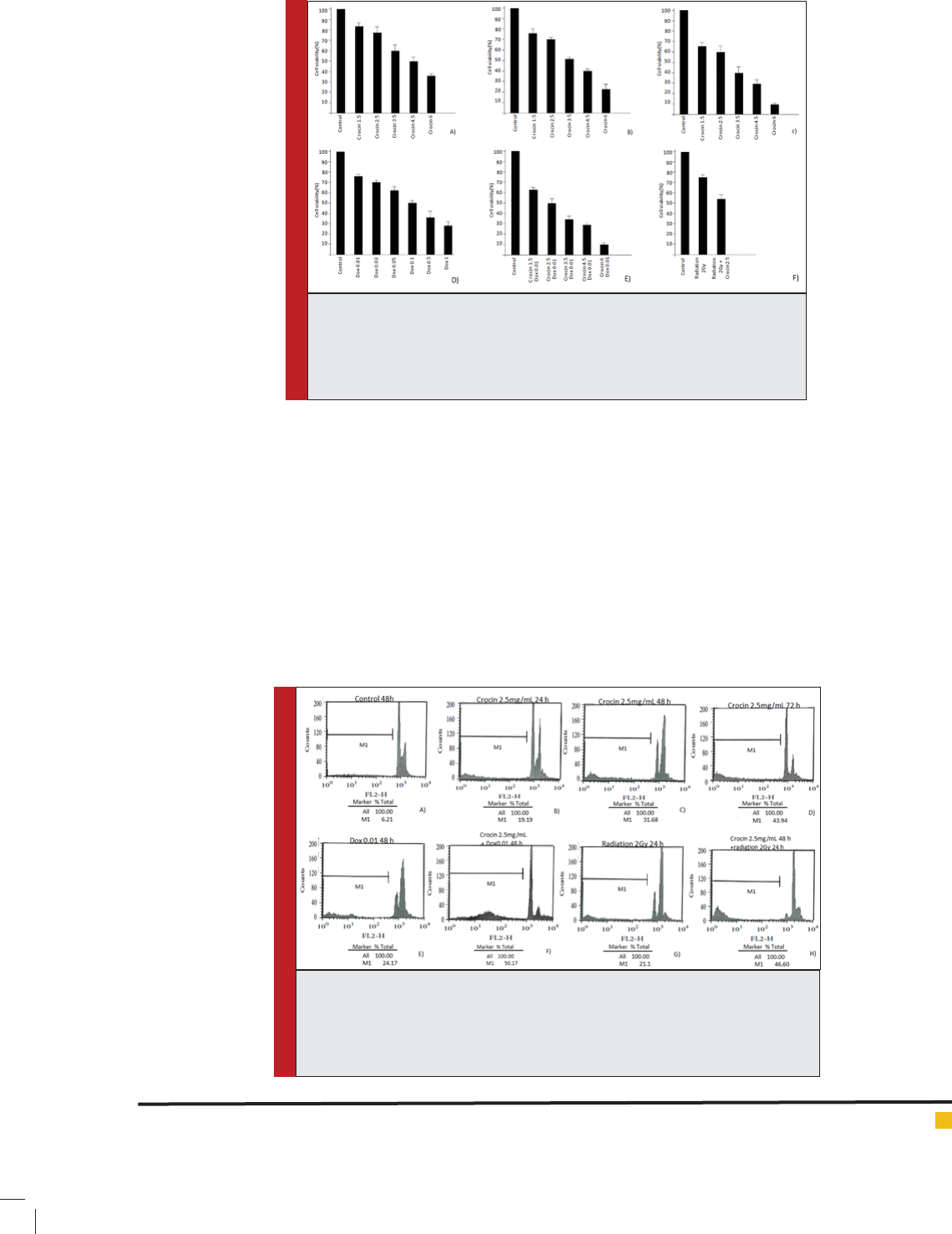
This study revealed the cell growth of MICHIGAN CAN-
CER FOUNDATION-7 cell line was inhibited by crocin in
a dose and time dependent manner (Figure 1 A-C). There
were signi cant differences among different groups (P
value <0.05). IC50 was shown for 3.5 mg/ml crocin in 48
h by MTT (Figure 1 B).The resultes of Crocin MTT asseys
are similar to results of Vali and changizi 2015.
Different concentrations of doxorubicin for 48 incu-
bation could suppress MICHIGAN CANCER FOUNDA-
TION-7 cell line in a dose dependent manner (P value
<0.05) (Fig.1D). For this drug IC50 was obtained 0.1 µM/
ml in 48 h by MTT assay. This research showed com-
bination of different concentrations of crocin and 0.01
µM/ml doxorubicin, may have stronger inhibition effect
than the single agent on breast cancer cells. Similarly it
was shown with combination of 2.5mg/ml of crocin and
430 EFFECT OF CROCIN AND DOXORUBICIN / RADIATION ON THE BREAST CANCER CELL LINE BIOSCIENCE BIOTECHNOLOGY RESEARCH COMMUNICATIONS
BIOSCIENCE BIOTECHNOLOGY RESEARCH COMMUNICATIONS EFFECT OF CROCIN AND DOXORUBICIN / RADIATION ON THE BREAST CANCER CELL LINE 431
Ali Reza Fanayi et al.
2Gy radiation the survival rate was signi cantly lower
(P value <0.05) (Fig. 1E _
APOPTOTIC CHANGES OF MICHIGAN CANCER
FOUNDATION-7 CELLS
Apoptosis following treatment with crocin, doxorubicin,
radiation and combination of them was measured by
owcytometry using PI staining to detect the sub-G1
peak resulting from DNA fragmentation. The results
showed apoptosis rate with crocin to be increased with
duration of time (P < 0.05) (Fig. 2 A _ D). Also this study
revealed the combined treatment of crocin and doxoru-
bicin or crocin and radiation have more apoptotic effect
than the single agent (P < 0.05) (Fig. 2 E _ H).
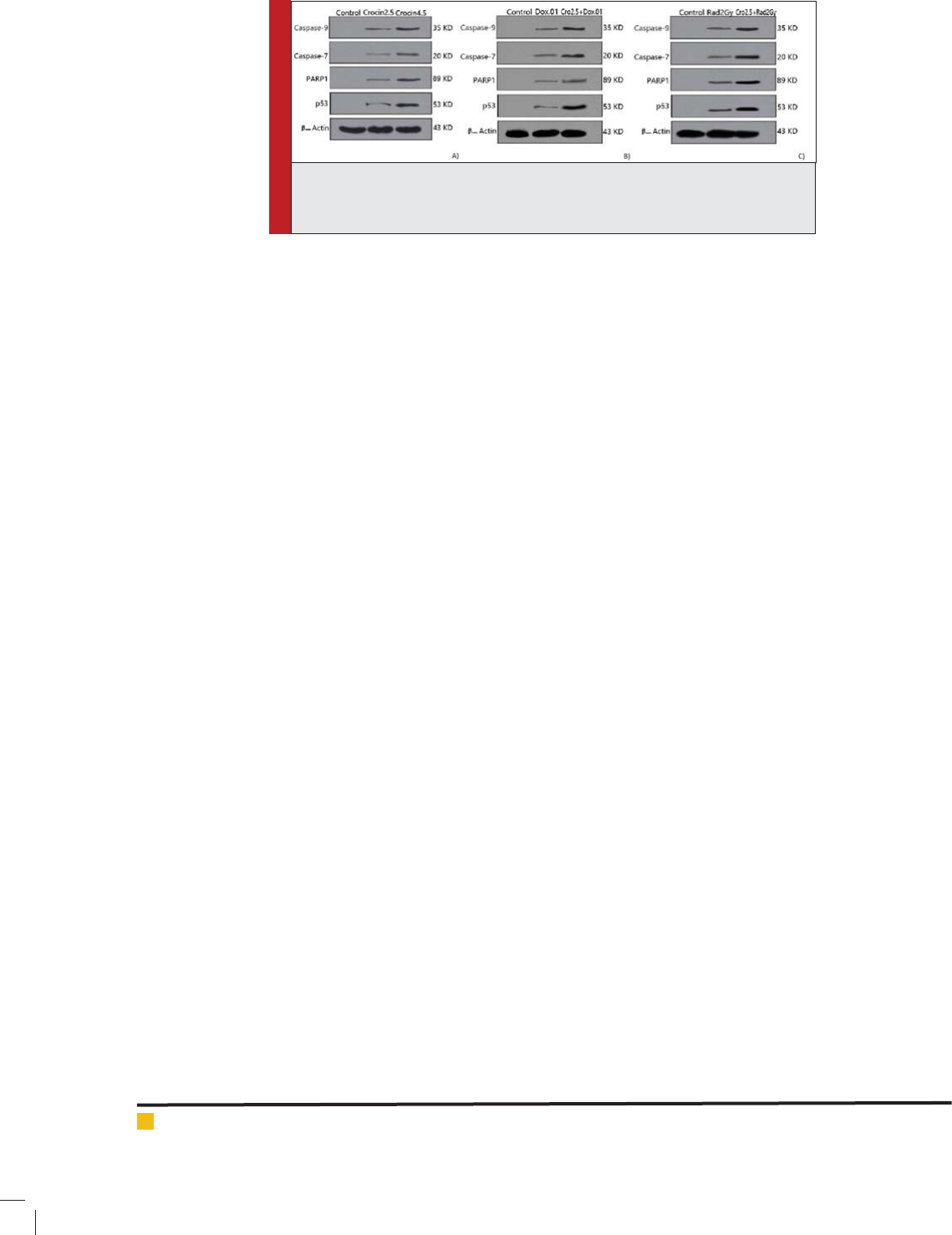
EXPRESSION OF CASPASE-9, CASPASE-7,
P53 AND PARP IN MICHIGAN CANCER
FOUNDATION-7 CELLS
With exposure of 2.5 mg/ml and 4.5 mg/ml crocin to
MICHIGAN CANCER FOUNDATION-7 cells for 48h,
FIGURE 1: The inhibition effect of crocin, Doxorubicin, Radiation and com-
bination of them on MICHIGAN CANCER FOUNDATION-7 cells, measured by
MTT (A) crocin 24h, (B) crocin 48h, (C) crocin 72 h. (D) Doxorubicin 48h (E)
combination of crocin in all concentration 48h with 0.01uM/ml Doxorubicin
48h (F) combination of crocin 48h with radiation 24h.
FIGURE 2: Flowcytometry histograms of PI-stained MICHIGAN CANCER FOUN-
DATION-7 cells and Sub-G1 peak means apoptotic cells.(A) control 48h (B)
Crocin 2.5 mg/ml 24h (C) Crocin 2.5 mg/ml 48h (D) crocin 2.5 mg/ml 72h (E)
doxorubicin .01uM/ml 48h (F) crocin 2.5mg/ml 48h + doxorubicin .01uM/ml (G)
radiation 2Gy (H) crocin 2.5 mg/ml 48h + radiation 2Gy 24h.
432 EFFECT OF CROCIN AND DOXORUBICIN / RADIATION ON THE BREAST CANCER CELL LINE BIOSCIENCE BIOTECHNOLOGY RESEARCH COMMUNICATIONS
Ali Reza Fanayi et al.
expression of p53, PARP, caspase-9 and caspase-7 was
detected. The results showed that the expression of all
four proteins was in a dose dependent manner (Fig. 3 A).
Also expression of these proteins was evaluated in com-
bination of crocin with doxorubicin or radiation. The
results showed that the expression of apoptotic proteins
in the combined treatments were signi cantly higher
than the single groups (Fig. 3 B_C).
DISCUSSION
Cancer is the second leading cause of death in the world.
In recent years its rate has been grown even more than
twice. Breast cancer is the most common cancer (23%
of all cancers) with the highest mortality (16%) among
all malignancies in women. Therefore breast cancer
remains one of the main health problem in the word
(Harirchi, Kolahdoozan et al. 2011). Studies show that
increased incidence of the breast cancer is higher in
developing countries and life expectancy of patients is
lower (Shibuya, Mathers et al. 2002). The reasons of can-
cer are associated with the environmental factors such
as air pollution, stress, diet and life style of the people. It
has been found that the consumption of foods that have
antioxidant properties, is effective in reducing the risk of
cancer (Ren, Qiao et al. 2003).
On the other hand, despite the use of therapeutic
options including surgery chemotherapy and radiother-
apy for patients mortality remains high. It indicates the
inef ciency of these ways. Moreover destructive effects
of chemotherapy and radiation on normal cell division
are also other disadvantages associated with the thera-
peutic process (Chabner Ba Fau - Friedman and Fried-
man). For example, while doxorubicin is a valuable anti-
cancer agent, cardiotoxicity and drug resistance are two
main problems of this drug (Kaye and Merry 1985, Weiss
1992).
Crocin is the main carotenoid of saffron with anti-
tumor properties (Jaliani, Riazi et al. 2013). Study
on HepG2 cell line showed telomerase activity in the
nucleus would decrease with increasing concentration
of crocin and it would lead to inhibition of prolifera-
tion and apoptosis (Noureini and Wink 2012). Our study
could approve this result. It was also found that DNA
fragmentation and cell cycle arrest are the main signs
of apoptosis in pancreatic cancer cells treated with
crocin(Bakshi, Sam et al. 2010). Gupta and colleagues in
October 2014 investigated the synergistic effect of crocin
and cisplatin on MDA MB-231 cells and MCF_7. It was
found that lower concentrations of cisplatin along with
crocin can achieve the desired result (Gupta, Jhamb et al.
2014). Notably, crocin signi cantly inhibits the growth
of cancer cells with no effects on normal cells (Sun, Xu
et al. 2013).
Consistent with the results of other studies, the results
of this study also con rms the effectiveness of crocin
against breast cancer Michigan center foundation-7 cell
line. To evaluate the survival rate of Michigan center
foundation-7 cells in treatment with doxorubicin, crocin,
radiation and combined therapy MTT assay was used.
This study showed treatment of Michigan center foun-
dation -7 cells with crocin was time and dose-dependent
to decrease the rate of proliferation. For example when
the concentration of crocin increased from 1.5 mg/ml to
6mg/ml in 48h, the survival rate of MCF-7 cells reduced
from 75% to 23%. Also the survival rate of MCF-7cells
reduced from 75% to 22% in treatment of cells with
doxorubicin from .01 µM/ml to 1µM/ml in 48h. It means
doxorubicin reduces the survival rate in a dose depend-
ent manner.
Cellular stresses such as ionizing radiation, UV and
chemical carcinogens can activate p53, including dam-
age to DNA, oncogene expression, hypoxia and nucleo-
tide depletion (Giaccia and Kastan 1998). According to
the type and severity of toxicity, p53 protein causes cell
cycle arrest or cell death through apoptosis that the for-
mer cause to repair DNA and the second led to remove
from cell replication(Bouvard, Zaitchouk et al. 2000) . P53
causes to release cytochrome C from mitochondria and
induces the expression of Apaf-1 forming the apopto-
FIGURE 3: Evaluating the expression of caspase-9, caspase-7, p53 and PARP (A)
in a concentration of 2.5 and 4.5 mg/ml in 48 h. (B) in dox .01uM/ml , crocin
2.5mg/ml + dox.01uM/ml (c) in radiation 2Gy , crocin2.5+radiation2G.

BIOSCIENCE BIOTECHNOLOGY RESEARCH COMMUNICATIONS EFFECT OF CROCIN AND DOXORUBICIN / RADIATION ON THE BREAST CANCER CELL LINE 433
Ali Reza Fanayi et al.
some (Kannan, Kaminski et al. 2001). Apoptosome leads
to activate caspase-9 following with executive caspases
such as 3,6 and 7 (Hengartner 2000). The caspase-acti-
vated DNase cut DNA between nucleosomes (Pecorino).
This process leads to measure DNA content after stain-
ing nucleic acid with speci c uorochromes(Riccardi
and Nicoletti 2006) . PI is a uorogenic compound that
binds to nucleic acids, so that uorescence emission is
proportional to the DNA content of a cell. When apop-
totic cells are stained with PI and analyzed with a ow
cytometer, they show a wide hypodiploid (sub-G1) peak,
that easily discriminates from narrow peak of normal
cells (diploid cells ) (Riccardi and Nicoletti 2006).
Michigan center foundation is a cancer cell line with
defect in caspase-3 and is relatively insensitive to many
chemotherapy drugs (Yang, Sladek et al. 2001). PARP
as the cellular protein is cleaved speci cally in apopto-
sis. Particular proteolysis of PARP happens in the DNA
binding domain. Caspase-3 and caspase-7 are the most
effective proteases for PARP cleavage (Herceg and Wang
2001). Therefore PI owcytometry and western blot were
used to evaluate breast cancer cells apoptosis in our study.
Flowcytometry showed to treat MCF 7cells for 48 hours
with 0.01 µmol/ml doxorubicin causes 24.17% apoptosis.
The combined therapy of 2.5 mg/ml crocin and 0.01µm/
ml Doxorubicin for 48 hours with a synergistic effect
caused 50.17% apoptosis. Also the combined therapy
of crocin and gamma radiation with a synergistic effect
could cause 46.60% apoptosis in breast cancer cells.
CONCLUSION
This study revealed the expression of apoptotic proteins
in the combined therapy of saffron and radiation or
saffron and drug was signi cantly higher than that in
using radiation or drug alone. Finally it was found that
crocin could be an appropriate supplement for treatment
of breast cancer by reducing the dosage and harmful
effects of drugs or radiation.
ACKNOWLEDGMENT
This study has been supported by Tehran University of
Medical Sciences. Grant number: 25349
CONFLICT OF INTEREST
There is no con ict of interest
REFERENCES
Baks hi, H., S. Sam, R. Rozati, P. Sultan, T. Islam, B. Rathore,
Z. Lone, M. Sharma, J. Triphati and R. C. Saxena (2010). DNA
fragmentation and cell cycle arrest: a hallmark of apoptosis
induced by crocin from kashmiri saffron in a human pancre-
atic cancer cell line. Asian Pac J Cancer Prev 11(3): 675-679.
Bouv ard, V., T. Zaitchouk, M. Vacher, A. Duthu, M. Canivet,
C. Choisy-Rossi, M. Nieruchalski and E. May (2000). Tissue
and cell-speci c expression of the p53-target genes: bax, fas,
mdm2 and waf1/p21, before and following ionising irradiation
in mice. Oncogene 19(5): 649-660.
Chab ner Ba Fau - Friedman, M. A. and M. A. Friedman Pro-
gress against rare and not-so-rare cancers.
Chry ssanthi, D. G., F. N. Lamari, G. Iatrou, A. Pylara, N. K.
Karamanos and P. Cordopatis (2007). Inhibition of breast can-
cer cell proliferation by style constituents of different Crocus
species. Anticancer Research 27(1A): 357362.
Cris well, T., K. Leskov, S. Miyamoto, G. Luo and D. A. Booth-
man (2003). Transcription factors activated in mammalian
cells after clinically relevant doses of ionizing radiation.Onco-
gene 22(37): 5813-5827.
Czec zuga-Semeniuk, E., S. Wołczynski, M. Dabrowska, J.
Dziecioł and T. Anchim (2004). The effect of doxorubicin and
retinoids on proliferation, necrosis and apoptosis in MICHI-
GAN CANCER FOUNDATION-7 breast cancer cells. Folia His-
tochemica et Cytobiologica 42(4): 221-227.
Fuji wara, A., T. Hoshino and J. W. Westley (1985). Anthracy-
cline antibiotics.Critical Reviews in Biotechnology 3(2): 133-
157.
Giac cia, A. J. and M. B. Kastan (1998). The complexity of p53
modulation: emerging patterns from divergent signals. Genes
& development 12(19): 2973-2983.
Gupt a, S., B. Jhamb and S. Katiyar (2014). Crocin-supple-
mented cisplatin is highly effective in killing breast cancer
cells than cisplatin alone.Cancer Research 74(19 Supplement):
4585-4585.
Hari rchi, I., S. Kolahdoozan, M. Karbakhsh, N. Chegini, S.
Mohseni, A. Montazeri, A. Momtahen, A. Kashe and M. Ebra-
himi (2011). Twenty years of breast cancer in Iran: downstag-
ing without a formal screening program.Annals of oncology
22(1): 93-97.
Heng artner, M. O. (2000). The biochemistry of apoptosis.
Nature 407(6805): 770-776.
Herc eg, Z. and Z.-Q. Wang (2001). Functions of poly (ADP-
ribose) polymerase (PARP) in DNA repair, genomic integrity
and cell death. Mutation Research/Fundamental and Molecular
Mechanisms of Mutagenesis 477(1): 97-110.
Jali ani, H. Z., G. H. Riazi, S. M. Ghaffari, O. Karima and A.
Rahmani (2013). The effect of the Crocus sativus L. carotenoid,
crocin, on the polymerization of microtubules, in vitro. Iranian
journal of basic medical sciences 16(1): 101.
Kann an, K., N. Kaminski, G. Rechavi, J. Jakob-Hirsch, N.
Amariglio and D. Givol (2001). DNA microarray analysis of
genes involved in p53 mediated apoptosis: activation of Apaf-
1.” Oncogene 20(26): 3449-3455.
Kaye , S. and S. Merry (1985). Tumour cell resistance to anthra-
cyclines—a review. Cancer chemotherapy and pharmacology
14(2): 96-103.

434 EFFECT OF CROCIN AND DOXORUBICIN / RADIATION ON THE BREAST CANCER CELL LINE BIOSCIENCE BIOTECHNOLOGY RESEARCH COMMUNICATIONS
Ali Reza Fanayi et al.
Li, X., T. Huang, G. Jiang, W. Gong, H. Qian and C. Zou (2013).
Synergistic apoptotic effect of crocin and cisplatin on osteo-
sarcoma cells via caspase induced apoptosis. Toxicology letters
221(3): 197-204.
Luzh na, L. and O. Kovalchuk (2010). Modulation of DNA
methylation levels sensitizes doxorubicin-resistant breast
adenocarcinoma cells to radiation-induced apoptosis. Bio-
chemical and biophysical research communications 392(2): 113-
117.
Møll er, P., H. Wallin and L. E. Knudsen (1996). Oxidative stress
associated with exercise, psychological stress and life-style
factors.Chemico-biological interactions 102(1): 17-36.
Nagh izadeh, B., M. T. Boroushaki, N. Vahdati Mashhadian and
S. M. T. Mansouri (2008). Protective effects of crocin against
cisplatin-induced acute renal failure and oxidative stress in
rats.Iranian Biomedical Journal 12(2): 93-100.
Nico letti, I., G. Migliorati, M. Pagliacci, F. Grignani and C. Ric-
cardi (1991). A rapid and simple method for measuring thymo-
cyte apoptosis by propidium iodide staining and ow cytom-
etry.” Journal of immunological methods 139(2): 271-279.
Nour eini, S. K. and M. Wink (2012). Antiproliferative effects
of crocin in HepG2 cells by telomerase inhibition and hTERT
down-regulation.Asian Pac J Cancer Prev 13(5): 2305-
2309.
Octa via, Y., C. G. Tocchetti, K. L. Gabrielson, S. Janssens, H. J.
Crijns and A. L. Moens (2012). Doxorubicin-induced cardiomy-
opathy: from molecular mechanisms to therapeutic strategies.
Journal of molecular and cellular cardiology 52(6): 1213-1225.
Peco rino, L. Molecular biology of cancer: mechanisms, targets,
and therapeutics, and therapeutics,3rd ed, United Kingdom:
Oxford university press; 2012 p152.
Ren, W., Z. Qiao, H. Wang, L. Zhu and L. Zhang (2003). Fla-
vonoids: promising anticancer agents. Medicinal research
reviews 23(4): 519-534.
Ricc ardi, C. and I. Nicoletti (2006). Analysis of apoptosis by
propidium iodide staining and ow cytometry. Nature proto-
cols 1(3): 1458-1461.
Schm idt, M., G. Betti and A. Hensel (2007). Saffron in phyto-
therapy: pharmacology and clinical uses. Wiener Medizinische
Wochenschrift 157(13-14): 315-319.
Sc ott, S. P. and T. K. Pandita (2006). The cellular control of
DNA doublestrand breaks.Journal of cellular biochemistry
99(6): 1463-1475.
Shib uya, K., C. D. Mathers, C. Boschi-Pinto, A. D. Lopez and C.
J. Murray (2002). Global and regional estimates of cancer mor-
tality and incidence by site: II. Results for the global burden of
disease 2000. BMC cancer 2(1): 37.
Stee l G, . B. C. R. L. -Sun, Y., H.-J. Xu, Y.-X. Zhao, L.-Z. Wang,
L.-R. Sun, Z. Wang and X.-F. Sun (2013). Crocin exhibits anti-
tumor effects on human leukemia HL-60 cells in vitro and in
vivo. Evidence-Based Complementary and Alternative Medi-
cine 2013.
Tazh ibi, M. and A. Feizi (2014). Awareness Levels about Breast
Cancer Risk Factors, Early Warning Signs, and Screening and
Therapeutic Approaches among Iranian Adult Women: A large
Population Based Study Using Latent Class Analysis.BioMed
research international 2014.
Vali,F.and Changizi,V(2015).Synergistic Apoptotic Effect of
Crocin and Paclitaxel or Crocin and Radiation on MCF-7 Cells,
a Type of Breast Cancer Cell Line.Intrenationali journal of
breast cancer 139349, 7page
Wei ss, R. B. (1992). The anthracyclines: will we ever nd a bet-
ter doxorubicin? Seminars in oncology.
Yang , X.-H., T. L. Sladek, X. Liu, B. R. Butler, C. J. Froelich
and A. D. Thor (2001). Reconstitution of caspase 3 sensitizes
MICHIGAN CANCER FOUNDATION-7 breast cancer cells to
doxorubicin-and etoposide-induced apoptosis.Cancer research
61(1): 348-354.