
Microbiological
Communication
Biosci. Biotech. Res. Comm. 9(3): 457-462 (2016)
Biotyping and antibiogram of
Riemerella anatipestifer
from ducks in Kerala
Surya, P. S.
1
, Priya, P. M.
2
* and Mini, M.
3
1
MVSc Scholar, Department of Veterinary Microbiology, College of Veterinary and Animal Sciences,
Mannuthy, Thrissur, Kerala-680651.
2
Assistant Professor, e of Veterinary and Animal Sciences, Mannuthy, Thrissur, Kerala-680651.
3
Professor and Head Department of Veterinary Microbiology, College of Veterinary and Animal Sciences,
Mannuthy, Thrissur, Kerala-680651.
ABSTRACT
New duck disease caused by Riemerella anatipestifer is a new disease emerged in Kerala from 2008 onwards. Six R.
anatipestifer isolates responsible for the disease were isolated from suspected ducks from different outbreak areas of
the state and were identi ed. Since the ecological, morphological and cultural characteristics of R. anatipestifer are
more or less similar to Pasteurella multocida, the disease is often confused with duck pasteurellosis and misdiagnosed.
R. anatipestifer infection is also characterized by the presence of bipolar organisms in blood smear and impression
smears of organs as in the case of P. multocida, but the size is little larger. The detection and identi cation of the
causative bacterium, from ducks with signs and lesions consistent with the acute or chronic form of the disease, is one
of the most important aspects of disease diagnosis. Hence, a study was conducted to isolate the agent of new duck
disease and stating its differential biotyping characters from that of P. multocida. They were differentiated using tests
like indole production, gelatin liquefaction, ornithine decarboxylases utilization and fermentation of glucose. The
antibiogram pattern was determined to advocate the choice of drug for the purpose of treatment. All the R. anatipes-
tifer isolates were sensitive to chloramphenicol, cipro oxacin, enro oxacin, nor oxacin, gentamicin, clindamycin,
doxycycline and cefuroxime.
KEY WORDS: ANTIBIOGRAM, BIOTYPING, KERALA, NEW DUCK DISEASE,
PASTEURELLA MULTOCIDA, RIEMERELLA ANATIPESTIFER
457
ARTICLE INFORMATION:
*Corresponding Author: priya@kvasu.ac.in
Received 18
th
July, 2016
Accepted after revision 7
th
Sep, 2016
BBRC Print ISSN: 0974-6455
Online ISSN: 2321-4007
Thomson Reuters ISI ESC and Crossref Indexed Journal
NAAS Journal Score 2015: 3.48 Cosmos IF : 4.006
© A Society of Science and Nature Publication, 2016. All rights
reserved.
Online Contents Available at: http//www.bbrc.in/

458 CHARACTERISATION OF
RIEMERELLA ANATIPESTIFER
BIOSCIENCE BIOTECHNOLOGY RESEARCH COMMUNICATIONS
Surya, Priya And Mini
INTRODUCTION
God’s gift of beautiful water bodies at various localities
of Kerala are acting as ideal environment for duck rear-
ing. Regular vaccination against duck plague and duck
pasteurellosis carried out in the state greatly reduced
their incidence. When we succeed in controlling the
existing disease, due to known and unknown global
environmental changes, several new diseases are emerg-
ing. One such disease is the new duck disease in Kerala,
reported since 2008 (Priya et al., 2008). It is an enzootic,
contagious, often primary septicemic disease of domes-
ticated ducklings (Fulton and Rimler, 2010).
In addition to ducks, it also infects geese, turkey,
chicken, wild birds and domestic pigs (Segers et al.,
1993). In young ducklings, it results in a mortality rate
as high as 75 per cent and in adult birds, it ranges from
20 to 40 per cent. The causative agent is Riemerella
anatipestifer, a Gram- negative rod shaped, non-motile,
non-sporulating bacterium.
In India, the disease has been reported in ducks
from Assam and Kerala (Shome et al., 2004 and Priya
et al.,2008). Both R. anatipestifer and Pasteurella mul-
tocida reveal bipolarity in blood smear on Lieshman’s
/ Giemsa staining.Both the organisms share common
ecological and morphological characters . Hence, the
eld veterinarians are often unable to distinguish these
two organisms due to their phenotypic similarity. Here
comes the need of isolation and identi cation of the
agent. The present study discussed in detail on direct
microscopic examination, right clinical samples to be
collected, selection of cultural media and its incubation
condition and the differential biochemical characters of
R. anatipestifer from that of P. multocida. These param-
eters are highly useful at eld level to con rm the dis-
ease, (Sun et al., 2012, Pala and Radhakrishnan 2014.,
Soman et al., 2014).
MATERIAL AND METHODS
Live and dead ducks (128) from the disease suspected
outbreak areas were brought to the Department of Vet-
erinary Microbiology was used for sample collection.
Detailed post mortem examination was conducted to
observe various gross lesions. Heart blood smears and
impression smears of liver and spleen were stained by
Leishman’s stain for the presence of bipolar organisms.
Samples of heart blood, liver, spleen, lungs and brain
were collected aseptically and streaked on ten per cent
bovine blood agar. They were incubated microaerophili-
cally in a candle jar at 37°C for 48 hours. The bacte-
rial isolates were identi ed based on morphological and
staining reactions, cultural and biochemical characters.
Since P. multocida is the most confusing organism with
R. anatipestifer, duck isolate of P. multocida serotype A
(maintaining in the department) was used for compari-
son as a negative control. Antibiotic sensitivity pattern
of the isolates was determined by standard disc diffusion
method (Bauer et al., 1966).
RESULTS AND DISCUSSION
Examination of heart blood smears and liver impression
smears revealed bipolar organisms which are relatively
larger in size than P. multocida, indicating the importance
of examination of heart blood and impression smears
from liver and spleen. Pillai et al. (1993) also noted the
size difference of bipolarity between these two organisms.
Since the clinical signs and gross lesions of new duck
disease are similar to diseases like duck pasteurellosis and
E. coli infection, the gold standard method of diagno-
sis is the isolation of the bacteria from clinical materials
in suitable media. So the isolation was tried from heart
blood, lung, liver, spleen, ovary and brain. In acute stage
of the disease, the organism could be readily isolated from
heart blood, liver, spleen, lungs and brain (Pathanasophon
et al., 1994). Bisgaard (1995) suggested that R. anatipesti-
fer often resulted in chronic salpingitis in surviving duck
and geese. So isolation was also tried from ovary of the
infected bird. According to Gooderham (1996), the best
source of isolation of the organism was brain.
In this study, though isolates were obtained mainly
from heart blood and liver, chance of contamination
was less in brain. The primary isolation was carried out
in ve to ten per cent bovine blood agar. According to
Rimler et al. (1998) no selective and/or indicative media
had been used for the isolation of R. anatipestifer and
the isolation of the organism from clinical materials
was sometimes dif cult due to the overgrowth of other
organisms (Higgins et al., 2000). Chocolate agar (Leavitt
and Ayroud, 1997), ovine blood agar (Crasta et al., 2002)
and ten per cent bovine blood agar (Priya et al. 2008
and Pala et al. 2014) have reported to be useful for the
primary isolation of R. anatipestifer.
The incubation carried out in a candle jar with mild
CO
2
tension at 37°C for 48 h was found to be optimum
for the culture of R. anatipestifer from clinical materials.
Smith et al. (1987) suggested that the organism preferred
microaerophilic environment for initial isolation. Segers
et al. (1993) reported that the organism grew best at
temperature 35 to 37°C after a primary isolation in a CO
2
enriched atmosphere. The ndings of the present study
are in agreement with the observations made by earlier
workers.
Following incubation of clinical samples in bovine
blood agar, convex, entire, transparent and butyrous
colonies suggestive of R. anatipestifer obtained from six
birds were designated as RA1 to RA6. These observa-
BIOSCIENCE BIOTECHNOLOGY RESEARCH COMMUNICATIONS CHARACTERISATION OF
RIEMERELLA ANATIPESTIFER
459
Surya, Priya And Mini
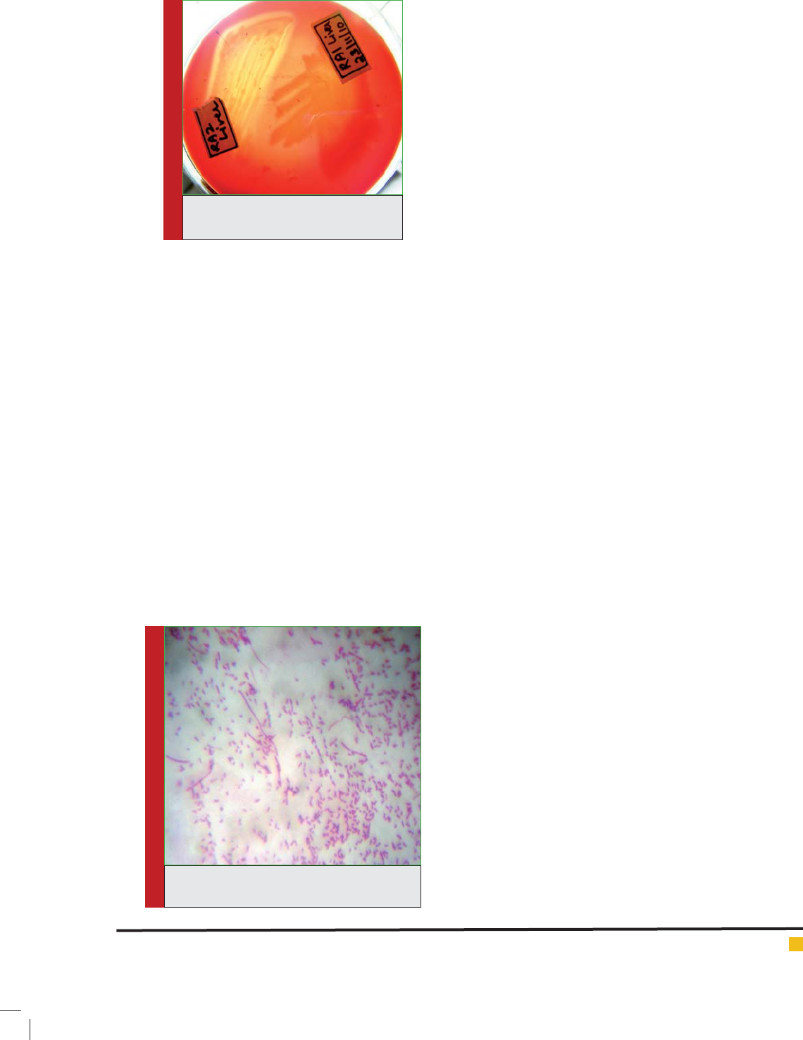
FIGURE 2. Variable morphology of R. anati-
pestifer
tions are in accordance with the ndings of Smith et al.
(1987) and Songer and Post (2005). All the isolates were
non-haemolytic on blood agar, except one (RA2), which
produced a clear zone of haemolysis after 48 h of incu-
bation (Fig. 1), indicating it may be a different strain
or serotype, since more than 20 serotypes of R. anati-
pestifer have been reported worldwide (Sandhu. 2008).
Hinz et al. (1998b) recorded that among 123 eld strains
of R. anatipestifer, 25 strains displayed haemolysis
on blood agar after 24 h to 48 h of incubation. In the
present study, out of the 128 birds screened, samples
from six birds were showing colonies suggestive of R.
anatipestifer.
Smears from culture, stained by Gram’s staining
revealed Gram negative organism with a variable mor-
phology varying from short rods to lamentous forms
(Fig. 2). Similar ndings were reported by Baba et al.
(1987) and Leavitt and Ayroud, (1997). The biochemical
characteristics of the isolates and its comparison with
DP1 are given in Table 1. The isolates did not grow on
MacConkey agar. They were catalase and oxidase posi-
tive and unreactive to O-F test, as reported by Carter
and Wise (2004) and Pala et al. (2013). According to
Hinz et al. (1998a) and Ryll et al. (2001), R. anatipestifer
is characterized more by the absence than the presence
of speci c phenotypic properties. The second stage bio-
chemical reactions used for characterization of R. anati-
pestifer (Segers et al., 1993) were almost identical for
all the six isolates. Variations were observed only in the
presence of urease and fermentation of sugars. Similar
ndings have been reported by Pillai et al. (1993), Van-
canneyt et al. (1999), Bernardet et al. (2002) and Shome
et al. (2004). According to OIE (2008), P. multocida and
R. anatipestifer could be differentiated using tests like
indole production, ornithine decarboxylase utilization
and gelatin liquefaction. On the basis of morphological,
cultural and biochemical characteristics, all the isolates
were identi ed as R. anatipestifer and were differenti-
ated from P. multocida.
Once the organism reaches the brain, chemotherapy
is of limited value. Hence, a variety of chemotherapeutic
agents have been used in the early stage of the disease
itself to treat the infection. As there is often a wide vari-
ation in the responsiveness of R. anatipestifer to these
agents, in vitro drug sensitivity testing is essential for
the selection of an appropriate drug in a given situation.
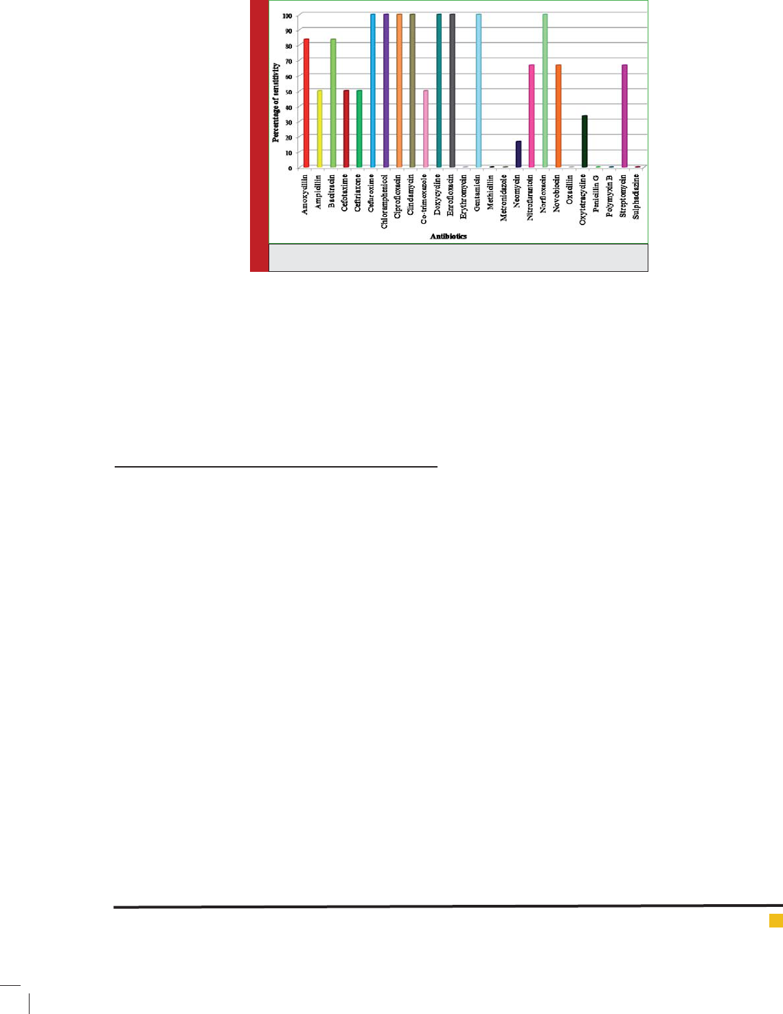
All the isolates used in the present study were subjected
to antibiotic sensitivity testing. Among the 26 antibiot-
ics used, cipro oxacin, enro oxacin, nor oxacin, doxy-
cycline, gentamicin, clindamycin, cefuroxime and chlo-
ramphenicol appeared to be the most effective drugs as
all the isolates tested were found to be sensitive to these
agents. Sensitivity to enro oxacin against R. anatipes-
tifer was reported by Turbahn et al. (1997). With regard
to the sensitivity of cipro oxacin, gentamicin, chlo-
ramphenicol and doxyxycline, the results of the present
study are in agreement with those of Shome et al. (2004)
and Priya et al. (2008).
All the isolates showed resistance to methicillin, met-
ronidazole, oxacillin, penicillin G, polymyxin B, eryth-
romycin and sulphadiazine. Other antibiotics tested
showed variable sensitivity pattern (Fig. 3). Several
workers reported the high sensitivity of R. anatipestifer
to penicillin G, erythromycin and polymyxin B (Baba et
al.,
1987; Pathanasophon et al., 1991 and Pathanasophon
et al., 1994). Chang et al.,(2003) conducted in vitro and
in vivo antibiogram using ceftiofur and 16 commonly
used antibiotics against 50 isolates of R. anatipestifer .
Their results revealed that penicillin, cephalothrin, cefti-
ofur, chloramphenicol, umequine and kanamycin are
the effective antibiotics.
In contrast to that, the study conducted by Zhong
et al., (2009) showed that the isolates were resistant
to penicillin, ampicillin, tetracycline and sensitive to
enro oxacin, chloramphenicol and neomycin.
FIGURE 1. Haemolysis produced by
RA2 on blood agar

460 CHARACTERISATION OF
RIEMERELLA ANATIPESTIFER
BIOSCIENCE BIOTECHNOLOGY RESEARCH COMMUNICATIONS
Surya, Priya And Mini
Table 1: Biochemical characteristics of Riemerella anatipestifer
TESTS RA1 RA2 RA3 RA4 RA5 RA6 DP1
Gram’s reaction - - - - - - -
Motility - - - - - - -
Growth microareobically - - - - - - -
Growth aerobically - - - - - - -
Growth on MacConkey agar - - - - - - -
Haemolysis on blood agar - + - - - - -
Catalase + + + + + + +
Oxidase + + + + + + +
O-F test - - - - - - F
Indole production - - - - - - +
Methyl-red test - - - - - - -
Voges-Proskauer test - - - - - - -
Urease + - - + + + -
H2S production - - - - - - -
Nitrate reduction - - - - - - +
Citrate utilization - - - - - - -
Gelatin liquefaction + + + + + + -
Ornithine decarboxylase - - - - - - +
Sugar fermentation
Dextrose - - - + + - +
Galactose - - - + - - +
Lactose - - - - + - -
Fructose - - - - + + +
Sucrose - + - + - + +
Xylose - - - - + - +
Mannose - - - - - - -
Maltose - - + - + + -
Mannitol - - - - - - +
Sorbitol - - - - - - +
Dulcitol - - - - - - -
Adonitol - - - - - - -
Inositol - - - - - - -
Salicin - - - - - - -
Inulin - - - - - - -
Arabinose - - - - - - +
Trehalose - - - + - + -
Melibiose - - - - - - -
Cellobiose - - - - - - -
Rhamnose - - - - - - -
Raf nose - + - - - - -
Zhang et al. (2014) stated the usefulness of levami-
zole as immunostimulant in the administration of adju-
vanated vaccine. Zhong et al. (2009) suggested that R.
anatipestifer drug resistance pro les changed over time.
So to reduce the irresponsible use of antibiotics, disc dif-
fusion analysis should be done for effective antibacterial
treatment.
Earlier, Sun et al. (2012) reported the prevalence of
multi- drug resistant R. anatipestifer isolates from China.
Manju et al., (2014) noticed that cipro oxacin, enro-
BIOSCIENCE BIOTECHNOLOGY RESEARCH COMMUNICATIONS CHARACTERISATION OF
RIEMERELLA ANATIPESTIFER
461
Surya, Priya And Mini
oxacin and gentamicin gave a wider zone of inhibition
where as the R. anatipestifer isolates tested were resist-
ant to amoxicillin, chloramphenicol and co-trimoxazole.
The variations in the antibiogram of the isolates in the
present study could be attributed to the indiscriminate
use of antibiotics either to treat the disease condition or
their increased use as feed additives, which might have
resulted in acquired drug resistance.
ACKNOWLEDGEMENTS
Authors are thankful to The Dean, College of Veterinary
and Animal Sciences, Mannuthy, Thrissur for providing
necessary facilities to carry out the work.
REFERENCES
Baba, T., Odagiri, Y., Morimoto, T., Horimoto, T. and Yamamoto,
S. (1987). An outbreak of Moraxella (Pasteurella) anatipestifer
infection in ducklings in Japan. Jpn. J. Vet. Sci. 49: 939-941.
Bauer, A.W., Kirby, W. M. M., Sherris, J. C. and Turck, M.
(1966). Antibiotic susceptibility testing by a standardized sin-
gle disk method. Am. J. Clin. Pathol. 45(4): 493-496.
Bernardet, J. F., Nagakawa, Y.and Holms, B. (2002). Proposed
minimal standards for describing new taxa of the family Fla-
vobacteriaceae and emended description of the family. Int. J.
Syst. Evol. Microbiol. 52: 1049-1070.
Bisgaard, M. (1995). Salpingitis in web-footed birds: preva-
lence; aetiology and signi cance. Avian Pathol. 24: 443-452.
Carter, G. R. and Wise, D. J. (2004). Essentials of Veterinary
Bacteriology and Mycology. (6
th
Ed.). Iowa State University
Press, USA. 290p.
Chang, C. F., Lin.W. H., Yeh, T. M., Chiang, T. S. and Chang, Y.
F. 2003. Antimicrobial susceptibility of Riemerella anatipesti-
fer isolated from ducks against the ef cacy of ceftriafur treat-
ment. J. Vet. Diagn. Invest. 15: 26-29.
Crasta, K. C., Chua, K. L., Subramaniam, S., Frey, J., Loh, H. and
Tan, H. M. (2002). Identi cation and characterization of CAMP
cohemolysin as a potential virulence factor of Riemerella
anatipestifer. J. Bacteriol. 184: 1932-1939.
Fulton, R. M. and Rimler, R. B. (2010). Epidemiologic investi-
gation of Riemerella anatipestifer in a commercial duck com-
pany by serotyping and DNA ngerprinting. Avian Dis. 54:
969-972.
Gooderham, K. R. (1996). Anatipestifer infection. In: Jordan, F.
T. W. and Pattinson, M. (eds.). Poultry Diseases. (4
th
Ed.). W. B.
Saunders Company Ltd. London. pp. 234.
Higgins, D. A., Henry, R. R. and Kounev, Z. V. (2000). Duck
immune response to Riemerella anatipestifer vaccines. Dev.
Comp. Immunol. 24: 153-167.
Hinz, K. H., Ryll, M. and Köhler, B. (1998a). Detection of acid
production from carbohydrates by Riemerella anatipestifer
and related organisms using the buffered single substrate test.
Vet. Microbiol. 60: 277-284.
Hinz, K. H., Ryll, M., Köhler, B. and Glünder, G. (1998b). Phe-
notypic characteristics of Riemerella anatipestifer and simi-
lar micro-organisms from various hosts. Avian Pathol. 27(1):
33-42.
Leavitt, S. and Ayroud, M. (1997). Riemerella anatipestifer
infection of domestic ducklings: Cross-Canada report. Can.
Vet. J. 38: 113.
OIE (The World Organization for Animal Health). (2008). Man-
ual of Diagnostic Tests and Vaccine for Terrestrial Animals. 1:
524-530.
Pala, S and Radhakrishnan, U. (2014). Genomic diversity of
Riemerella anatipestifer associated with outbreaks of new duck
disease in India. Indian J. Anim. Sci. 84 (11): 1166-1170.
Pala, S., Nair, U. R., Somu, C. and Mahendran,M. (2013). Molec-
ular diagnosisof new duck disease in India by 16 S rRNAgene
based PCR. Adv. Anim.Vet. Sci. 1 (5): 140=142.
Pathanasophon, P., Sawada, T. and Tanticharoenyos, T. (1991).
Characteristics of antimicrobial susceptabilities of Pasteurella
FIGURE 3. Antibiotic sensitivity pattern of R. anatipestifer isolates

462 CHARACTERISATION OF
RIEMERELLA ANATIPESTIFER
BIOSCIENCE BIOTECHNOLOGY RESEARCH COMMUNICATIONS
Surya, Priya And Mini
(Moraxella) anatipestifer isolated from ducks in Thailand. Thai.
J. Hlth. Resch. 5: 55-61.
Pathanasophon, P., Sawada, T. and Tanticharoenyos, T. (1994).
Physiologic characteristics, antimicrobial susceptibility and
serotypes of Pasteurella anatipestifer isolated from ducks in
Thailand. Vet. Microbiol. 39: 179-185.
Pillai, R. M., James, P.C., Punnose, K. T., Sulochana, S.,
Jayaprakasan, V. and Nair, G. K. (1993). Outbreak of pasteurel-
losis among duck population in Kerala. J. Vet. Anim. Sci. 24:
34-39.
Priya, P. M., Pillai, D. S., Balusamy, C., Rameshkumar, P. and
Senthamilselvan, P. (2008). Studies on outbreak of new duck
disease in Kerela, India. Int. J. Poult. Sci. 7(2): 189-190.
Rimler, R. B., Sandhu, T. S. and Glisson, J. R. (1998). Rie-
merella anatipestifer infection. In: Swayne, D. E., Glisson, J.
R., Jackwood, M. H., Pearson, J. E. and Reed, W.M. (eds.).
The Laboratory Manual for the Isolation and Identi cation of
Avian Pathogens (4
th
Ed.). The American Association of Avian
Pathologists, Kennett square. pp. 22-23.
Ryll, M., Christensen, H., Bisgaard, M., Christensen, J. P.,
Hinz, K. H. and Kohler. B. (2001). Studies on the prevalence
of Riemerella anatipestifer in the upper respiratory tract of
clinically healthy ducklings and characterization of untypable
strains. J. Vet. Med. B. Infect Dis Vet Public Health. 48: 537-
546.
Sandhu, T. S. (2008). Riemerella anatipestifer infection. In:
Saif, Y. M., Fadly, A.M., Glisson,J. R., Mc Dougald, L. R., Nolan,
L.K. and Swayne, D. E. Diseases of Poultry (12
th
Ed.). Blackwell
publishing Professional, USA. pp. 758-764.
Segers, P., Mannheim,W., Vancanneyt, M., DeBrandt, K., Hinz,
K.H., Kersters, K. and Vandamme, P. (1993). Riemerella anati-
pestifer gen. nov., comp. nov., the causative agent of septice-
mia anserum exudativa, and its phylogenetic af liation within
the Flavobacterium-Cytophaga rRNA homology group. Int. J.
Syst. Bacteriol. 43: 768-776.
Shome, R., Shome, B. R., Rahman, H., Murugkar, H. V., Kumar,
A., Bhatt, B. P. and Bujarbarauah, K. M. (2004). An outbreak of
Riemerella anatipestifer infection in ducks in Meghalaya.
Indian J. Comp. Microbiol. Immunol. Infect. Dis. 25: 126-127.
Smith, J. D., Frame, D. D., Cooper, G., Bickford, A.A., Ghazikha-
nian, G. Y. and Kelly, B. J. (1987). Pasteurella anatipestifer
infection in commercial meat type turkeys in California. Avian
Dis. 31: 913-917.
Soman, M., Nair, S. R., Mini, M., Mani, B. K. and Joseph, S.
(2014). Isolation and polymerase chain reaction-based identi-
cation of Riemerella anatipestifer from duck in Kerala, India,
Vet. World . 7: 765-769.
Songer, J. E. and Post, K. W. (2005). Veterinary Microbiology:
Bacterial and Fungal Agents of Animal Disease. Elsevier Saun-
ders, Missouri. 434p.
Sun, N.,Liu, J. H., Yang, F., Lin, D., Li, G.,Chen, Z. and Zeng, Z.
(2012). Molecular characterization of antimicrobial resistance
of Riemerella anatipestifer isolated from ducks. Vet.Microbiol.,
158 (3-4):376-383.
Turbahn, A., Jackel, S. C. D., Greuel, E., Jong, A. D., Froyman,
R. and Kaleta, E. F. (1997). Dose response study of enro oxacin
against Riemerella anatipestifer septicemia in Muscovy and
pekin ducklings. Avian Pathol. 26(4): 791-802.
Vancanneyt, M., Segers, P., Hauben, L., Hommez, J., Devriese,
L. A., Hoste, B., Vandamme, P. and Kersters, K. (1994). Fla-
vobacterium meningosepticum, a pathogen in birds. J. Clin.
Microbiol. 32: 2398-2403.
Zhang, Y., Chen, H.,Zeng, X., Wang, P., Li, J. and Wu, W.
(2014). Levamizole enhances immunity in ducklings vacci-
nated against Riemerella anatipestifer. Microbiol.
Immunol. 58 (8): 456-62.
Zhong, C. Y., Cheng, A. C., Wang, M. S., Zhu, D. K., Luo, Q. H.,
Zhong, C.D., Li, L. and Duan, Z. (2009). Antibiotic susceptibility
of Riemerella anatipestifer eld isolates. Avian Dis. 53: 601-607.