
Biomedical
Communication
Biosci. Biotech. Res. Comm. 9(3): 503-511 (2016)
Computational analysis of polymorphisms of ubiquitin
carboxyl–terminal esterase L1 (UCHL1) gene involved
in Parkinson’s disease
Sowmya Dhawan
1
* and Usha Chouhan
2
Department of Bioinformatics, Maulana Azad National Institute of Technology, Bhopal, M.P. 452051, India
ABSTRACT
Presence of genetic variations is a key player among many others which affect susceptibility and progression of the
disease. Single nucleotide polymorphisms are the most frequent variations in human genome. Ubiquitin carboxyl-
terminal esterase L1 (UCHL1) located on chromosome 4p14 is one of the potential candidate neuropathogenic pro-
tein involved in Parkinson’s Disease. The aim of this study was to investigate the functional consequences of UCHL
1 single nucleotide polymorphisms (SNPs) to understand the biological basis of complex traits and diseases as the
Genetics of human phenotypic variation could be understood by knowing the functions of SNPs derived from the
data available in dbsSNP data base and different computer applications are used. Nonsynymous SNPs are relevant
in many of the human inherited disease since they change the aminoacid sequence of the protein. Few common
single –nucleotide polymorphisms (SNPs) of the UCHL1 genes were analyzed by using different bioinformatics tools
based on evolutionary analysis- sequence homology based, structure based approach. Protein structural analysis was
also performed by using I- Mutant. It was recognized that rs6063 and rs74315205 SNPs of UCHL1 gene were found
to be more damaging in PD and is responsible for the alteration in the levels of expression. Conclusion: It has been
concluded that among the entire SNPs of UCHL1 gene, the mutation in rs6063 and rs74315205 have the most sig-
ni cant effect on functional variation. The study suggested that G191R, G199 R, G88R and R231G variants of UCHL1
could directly or indirectly destabilize the amino acid interactions and hydrogen bond networks thus explaining the
functional deviations of protein to some extent. These results may further form the basis of large- scale population
based association studies.
KEY WORDS: PARKINSON’S DISEASE, SINGLE NUCLEOTIDE POLYMORHISMS, SNP, UCHL 1 GENE
503
ARTICLE INFORMATION:
*Corresponding Author: sowmyadhawan@gmail.com
Received 30
th
July, 2016
Accepted after revision 14
th
Sep, 2016
BBRC Print ISSN: 0974-6455
Online ISSN: 2321-4007
Thomson Reuters ISI ESC and Crossref Indexed Journal
NAAS Journal Score 2015: 3.48 Cosmos IF : 4.006
© A Society of Science and Nature Publication, 2016. All rights
reserved.
Online Contents Available at: http//www.bbrc.in/

504 IDENTIFICATION OF NOVEL MICRO RNAS AND THEIR TARGETS BIOSCIENCE BIOTECHNOLOGY RESEARCH COMMUNICATIONS
Sowmya Dhawan and Usha Chouhan
INTRODUCTION
Parkinson’s disease (PD) is the second most common
neurodegenerative disorder after Alzheimer’s affecting
approximately 1–2% of the population over the age of
65 and reaching a prevalence of almost 4% in those aged
above 85. Resting tremor, bradykinesia, rigidity, and
postural instability are the main clinical symptoms of
the disease often accompanied by nonmotor symptoms
including autonomic insuf ciency, cognitive impair-
ment, and sleep disorders (Gómezet al.2015).
There are two forms of the disease, the sporadic and
familial forms. The patients with familial PD are dis-
tinguished from the ones who suffer from sporadic PD
because of the early onset, greater consanguinity rate,
and greater frequency of similar disease in their parents
Familial PD cases are of 10% of the total no of cases
and are based on the genetic component of the disease
(Christine and Ana 2012). There is a life risk of 1.3% for
women and 2% for men as per the study of Olmstead
country. The disease is going to increase in the future
to come due to the medical expenses and other reasons
(Prasad et al.2016).
UCHL1 /Park 5 gene is a compelling candidate gene
for PD (Maraganore et al.2004) on biological grounds
because the protein it encodes plays a pivotal role in
the ubiquitin proteasome system (UPS), displays neuron-
speci c expression and is found in Lewy bodies, the neu-
ropathologic hallmark of PD .The ubiquitin proteasome
system regulates the degradation of key regulatory pro-
teins as well as misfolded and damaged proteins (Aaron
& Yong 2014). Ubiquitin carboxy-terminal hydrolase L1
(UCHL1) is a 223-a.a. protein which is a component of
the UPS, which cleaves the carboxy-terminal peptide
bond of polyubiquitine chains, working as a deubiqui-
tinating enzyme (Liu et al.2002). It encodes for one of
the most abundant proteins in the brain. Mutations in
this target were found to be responsible for a genetic
form of PD. It is thought a mutation at amino acid posi-
tion 93 for methionine may decrease UCHL1 hydrolase
activity, leading to accumulation of proteins that should
have been degraded, and subsequently the progression
of PD (Contu et al.2014).
One of the important gene speci c mutations
described for the familial forms of PD, include autoso-
mal dominant mutations of UCHL1 (PARK5). However,
the pathogenic mechanisms underlying mitochondrial
dysfunction in familial PD require further detailed inves-
tigation at the molecular level. The loss of dopaminergic
neurons in PD is preceded by the formation of Lewy
Bodies, insoluble proteinaceous inclusions enriched
with ubiquitinated aggregates, as well as displaying
extensive protein oxidative modi cation, (Hyo and Sun
2015).
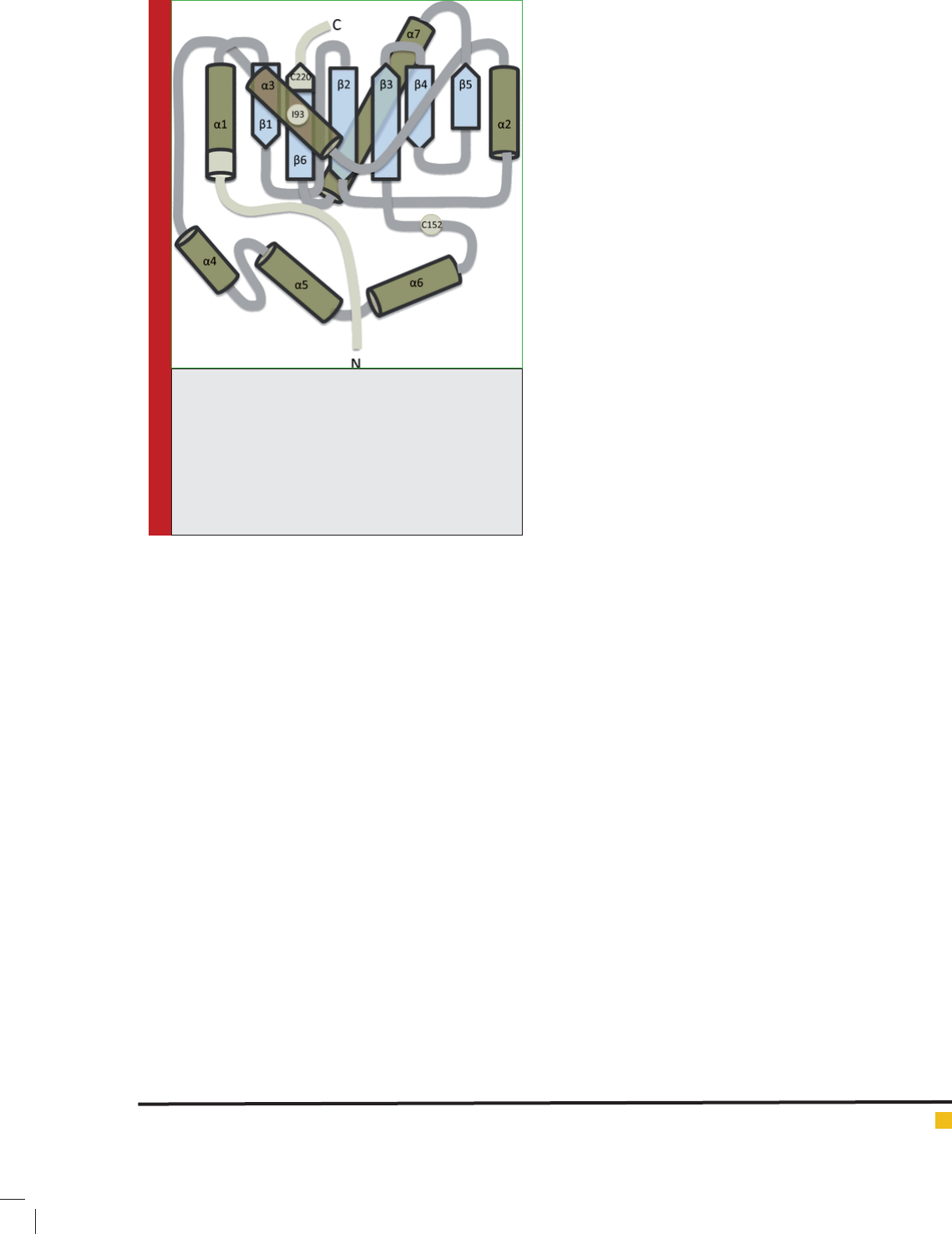
The structure of UCH-L1 contains a central -sheet
that is anked on either side by -helices as shown in
gure 1. In the crystal structure, UCH-L1 is an asymmet-
ric dimer; however, equilibrium sedimentation analysis
showed that the protein is monomeric in solution. The
catalytic triad comprises Cys90, His161, and Asp176; in
the crystal structure, the side chains of these residues are
not close enough for catalytic activity, suggesting that
in the absence of substrate, UCH-L1 is in an inactive
form. In addition, the active site is covered by a loop (L8)
that has been suggested to restrict the size of substrates
that can access the active-site cleft ( Bishop et al.2016).
A single nucleotide polymorphism (SNP) is a source
of variance in a genome. A SNP is a single base muta-
tion in DNA. SNPs are the most simple form and most
common source of individual genetic polymorphism
in the human genome (90% of human DNA polymor-
phisms). A SNP in a coding region may have two dif-
ferent effects on the resulting protein: Synonymous, the
substitution causes no amino acid change to the protein
it produces; non synonymous, the substitution results
in an alteration of the encoded amino acid. One half of
all coding sequence SNPs result in non- synonymous
codon changes (Smith 2002). A non- synonymous single
nucleotide polymorphism (nsSNP) occurring in a cod-
ing gene may cause an amino acid substitution in the
corresponding protein product, thus affecting the phe-
notype of the host organism .Non synonymous variants
constitute more than 50% of the mutations known to be
involved in human inherited diseases Single nucleotide
polymorphisms (SNPs) (Kumar 2009). Computational
methods are suf ciently fast and exible to provide reli-
able predictions of functionally signi cant SNPs with a
high accuracy of 80–85%when combined with sequence,
structure, and phylogenetic relationships (Minyue et al.,
2014). Here we are trying to consider computationally
a suitable protocol for missense mutation (point muta-
tion/single amino acid polymorphism) analysis before
wet lab experimentation and provided an optimal path
for further clinical and experimental studies.
MATERIAL AND METHODS
The data on protein sequence and variants (single
amino acid polymorphisms/missense mutations/point
mutations) for UCHL1 gene were collected from NCBI
database (http://www.ncbi.nlm.nih.gov/snp/)of SNP by
applying appropriate limits like homo-sapiens, Chromo-
some 4, cited in Pubmed etc. to detect the detrimental
point mutants.
Further deleterious SNP analysis were performed
using the computational tools sorting intolerant from
tolerant (SIFT) and Polyphen 2 for nsSNPs and FASTSNP
and UTRscan for UTR SNPs.
BIOSCIENCE BIOTECHNOLOGY RESEARCH COMMUNICATIONS IDENTIFICATION OF NOVEL MICRO RNAS AND THEIR TARGETS 505
Sowmya Dhawan and Usha Chouhan
SEQUENCE HOMOLOGY BASED METHOD (SIFT)
We have used the program SIFT (http://sift.bii.a-star.edu.
sg/index.html) to detect deleterious coding nonsynony-
mous SNPs. SIFT is a sequence homology-based tool to
predict whether an amino acid substitution in a protein
would be tolerated or damaging (Pauline et al., 2003). We
performed SIFT by submitting the query in the form of
SNP IDs or chromosome positions and alleles in nsSNVs
tool. Variants at the position with tolerance index score
#0.05 were considered as deleterious. A lower tolerance
index indicates that the particular amino acid substitution
likely has a more functional impact (Pauline et al., 2001).
STRUCTURE HOMOLOGY BASED METHOD
(POLYPHEN)
Analyzing the damaged coding nonsynonymous SNPs
at the structural level is considered to be very important
to understand the functional activity of the protein of
concern. We have used PolyPhen server (http://genet-
ics.bwh.harvard.edu/pph2/) for this purpose. This is an
automatic tool that predicts the possible impact of an
amino acid substitution on a number of features, includ-
ing the sequence, phylogenetic, and structural informa-
tion. The query was submitted in the form of protein
sequence with mutational position and substitution. The
PolyPhen output comprises a score that ranges from 0
to 1, with zero indicating a neutral effect of amino acid
substitutions on protein function. Conversely, a high
score represents a variant that is more likely to be dam-
aging (Ramensky et al., 2002).
FUNCTIONAL SIGNIFICANCE OF NONCODING
SNPS IN REGULATORY UNTRANSLATED
REGIONS
The Web server FastSNP (http://fastsnp.ibms.sinica.
edu.tw) was used for predicting the functional signi -
cance of the 5’ and 3’UTRs of the UCHL 1 gene (Hsiang
et al., 2006). The FastSNP server follows the decision
tree principle with external Web service access to TF
Search, which predicts whether a noncoding SNP alters
the transcription factor-binding site of a gene. The score
was given by this server on the basis of levels of risk
with a ranking of 0, 1, 2, 3, 4, or 5. This signi es the
levels of no, very low, low, medium, high, and very high
effect, respectively.
SCANNING OF UTR SNPS IN UTR SITE
The 5’ and 3’ UTRs are involved in various biologi-
cal processes such as posttranscriptional regulatory
pathways, stability, and translational ef ciency. We
used the program UTRscan (http://itbtools.ba.itb.cnr.it/
utrscan) which allows one to search the user-submitted
sequences for any of the patterns collected in the UTR
site (Graziano and Sabino 1999). UTRsite is a collection
of functional sequence patterns located in 5’ or 3’UTR
sequences. Brie y, two or three sequences of each UTR
SNP that have a different nucleotide at an SNP position
are analyzed by UTRscan, which looks for UTR func-
tional elements by searching through user-submitted
sequence data for the patterns de ned in the UTRsite
and UTR databases. If different sequences for each UTR
SNP are found to have different functional patterns, this
UTR SNP is predicted to have functional signi cance.
The Internet resources for UTR analysis are UTRdb and
UTRsite. UTRdb contains experimentally proven biologi-
cal activity of functional patterns of UTR sequence from
eukaryotic mRNAs(Graziano et al., 2002). The UTRsite
has the data collected from UTRdb and also is continu-
ously enriched with new functional patterns.
SUPPORT VECTOR MACHINE (I-MUTANT 3.0
AND FOLD- X)
The analyses were also conducted by using I-Mutant
Suite is a suite of support vector machine (SVM)- based
predictors of protein stability changes according to
Gibbs free energy change, enthalpy change, heat capac-
FIGURE 1. Schematic of UCH-L1 structure, Sche-
matic illustrating the -helical and -strand
structure of UCH-L1. The residues 1–11 at the
N-terminus, 220–223 at the C-terminus and residues
Ile
93
and Cys
152
are highlighted. It has been proposed
that modi cation at these points can affect the
hydrophobic core of -strands that are otherwise
protected from solution.
506 IDENTIFICATION OF NOVEL MICRO RNAS AND THEIR TARGETS BIOSCIENCE BIOTECHNOLOGY RESEARCH COMMUNICATIONS
Sowmya Dhawan and Usha Chouhan
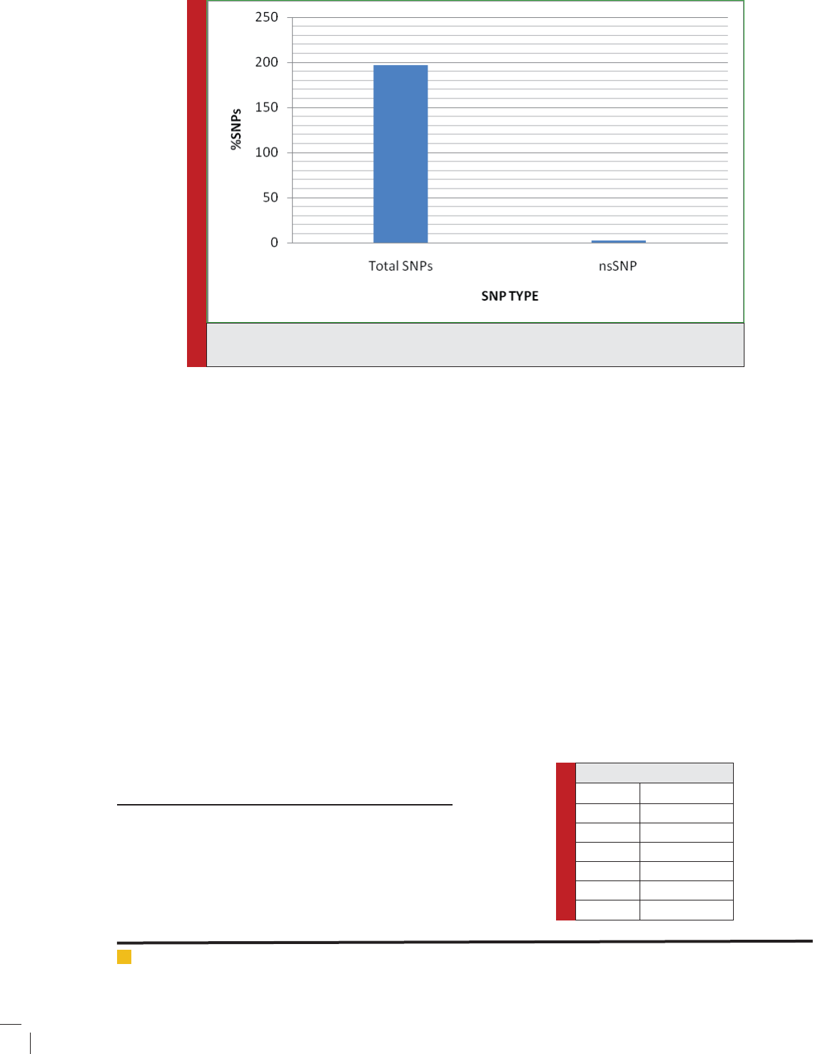
FIGURE 2. A graphical representation of distribution of nonsynonymous, SNPs for UCHL 1
(based on the dbSNP database).
ity change, and transition temperature (Capriotti et al.,
2005).The analysis was performed based on protein
sequence combined with mutational position and corre-
lated new residue. And the output result of the predicted
free energy change (DDG) classi es the prediction into
one of three classes: largely unstable (DDG, 20.5 kcal/
mol), largely stable (DDG.0.5 kcal/mol), or neutral (-0.5#
DDG#0.5 kcal/mol). IMutant Suite is available at (http://
gpcr2.biocomp.unibo.it/cgi/predictors/I-Mutant3.0/I-
Mutant3.0.cgi).
The FASTA sequence of protein retrieved from Uni-
Prot was used as an input to predict the mutational
effect on protein stability. I-Mutant also provides the
scores for free energy alterations, calculated with the
FOLD-X energy based web server (Schymkowitz et al.,
2005). FOLD-X is a computer algorithm for quantita-
tive estimation of interactions facilitating the stability
of proteins. The FOLD-X tool was used to provide the
comparison between wild type and mutant models in the
form of van der Waals clashes, which greatly in uence
the energy decomposition.
RESULTS AND DISCUSSION
SINGLE AMINO ACID POLYMORPHISM
DATASET FROM NCBI DBSNP DATABASE
The dbSNP database contains both validated and non-
validated polymorphisms. In spite of this drawback, we
opted to avail the dbSNP because the allelic frequency of
most of nsSNPs of UCHL 1 has been recorded there and
that is the most extensive SNP database. We selected 15
SNPs, out of which 2 were nsSNPs, as shown in Fig. 2.
DELETERIOUS SINGLE POINT MUTANTS
IDENTIFIED BY THE SIFT PROGRAM
The conservation level of a particular position in a protein
was determined by using a sequence homology-based
tool, SIFT. The protein sequences of 64 variants were sub-
mitted independently to the SIFT program to determine
the tolerance index. The higher the tolerance index, the
less functional impact a particular amino acid substitu-
tion is likely to have, and vice versa. Among the 64vari-
ants, 24 were found to be deleterious, having a tolerance
index score of ≤0.05. The results are shown in Table 2.
UTRSCAN ANALYSIS
Functional SNPs in UTR found by the UTRscan server
Polymorphisms in the 3’ UTR affect gene expression by
Table 1: SIFT classi cation
Ranking Risk Division
0 No effect
1 Very low
2 Low
3 Medium
4 High
5 Very high

BIOSCIENCE BIOTECHNOLOGY RESEARCH COMMUNICATIONS IDENTIFICATION OF NOVEL MICRO RNAS AND THEIR TARGETS 507
Sowmya Dhawan and Usha Chouhan
Table 2: SIFT analysis of SNPs, Variants with tolerance
index ≤0.05 score are considered as deleterious while
others are taken to be tolerant.
SNPs Amino acid
Change
Score Prediction
rs6063 G191R 0.002 DELETERIOUS
G191R 0.002 DELETERIOUS
G199R 0.002 DELETERIOUS
G199R 0.002 DELETERIOUS
G88R 0.003 DELETERIOUS
G88R 0.003 DELETERIOUS
rs1799895 R231G 0.017 DELETERIOUS
rs45454496 E3931K 0.037 DELETERIOUS
E3898K 0.037 DELETERIOUS
E941K 0.055 TOLERATED
E22K 0.057 TOLERATED
E1022K 0.147 DELETERIOUS
E1837K 0.186 TOLERATED
E1846K 0.188 TOLERATED
E529K 0.233 TOLERATED
E878K 0.368 TOLERATED
rs62625014 S389F 0.054 TOLERATED
S320F 0.101 TOLERATED
S320F 0.101 TOLERATED
S320F 0.101 TOLERATED
S320F 0.101 TOLERATED
rs63749888 E47Q 0.102 TOLERATED
E37Q 0.248 TOLERATED
rs66785829 V3601D 0.011 DELETERIOUS
V3634D 0.012 DELETERIOUS
V644D 0.063 TOLERATED
V201D 0.067 TOLERATED
V725D 0.193 TOLERATED
V550D 0.209 TOLERATED
V1549D 0.28 TOLERATED
V1540D 0.282 TOLERATED
rs74315205 E864K 0 DELETERIOUS
rs75353611 D25V 0.003 DELETERIOUS
D25V 0.003 DELETERIOUS
D27V 0.003 DELETERIOUS
D25V 0.003 DELETERIOUS
D25V 0.027 DELETERIOUS
rs112534524 G261A 0.27 TOLERATED
G261A 0.274 TOLERATED
G261A 0.277 TOLERATED
G261D 0.114 TOLERATED
G261D 0.119 TOLERATED
G261D 0.126 TOLERATED
rs121912705 T754N 0.039 DELETERIOUS
T3744N 0.053 TOLERATED
T3711N 0.055 TOLERATED
T311N 0.302 TOLERATED
T660N 0.386 TOLERATED
T835N 0.45 TOLERATED
T1659N 0.535 TOLERATED
T1650N 0.556 TOLERATED
rs121912706 R3873W 0.001 DELETERIOUS
R3906W 0.001 DELETERIOUS
R916W 0.002 DELETERIOUS
R1821W 0.003 DELETERIOUS
R997W 0.003 DELETERIOUS
R1812W 0.004 DELETERIOUS
R853W 0.049 DELETERIOUS
R504W 0.06 TOLERATED
rs180843436 E137K 0.014 DELETERIOUS
E486K 0.015 DELETERIOUS
E3537K 0.021 DELETERIOUS
E3570K 0.021 DELETERIOUS
E580K 0.06 TOLERATED
E661K 0.061 TOLERATED
E1485K 0.062 TOLERATED
E1476K 0.063 TOLERATED
rs199473343 L1622M 0.168 TOLERATED
L1655M 0.169 TOLERATED
T854N 0.105 TOLERATED
T3844N 0.147 TOLERATED
T3811N 0.148 TOLERATED
T411N 0.267 TOLERATED
T935N 0.356 TOLERATED
T1759N 0.432 TOLERATED
T1750N 0.434 TOLERATED
T760N 0.476 TOLERATED
rs386833750 CC2D2A 0 DELETERIOUS
rs386833752 T1065M 0.001 DELETERIOUS
T1114M 0.001 DELETERIOUS
affecting the ribosomal translation of mRNA or by in u-
encing the RNA half-life. Table 3 shows the list of SNPs
in the 3 that are predicted to be damaging because of the
presence of regulatory elements and are of functional
signi cance. We used the UTRscan server for this pur-
pose. We analyzed the same 64 variants in UTRscan that
were analyzed by the SIFT. The UTRscan server nds
patterns of regulatory region motifs from the UTR data-

Sowmya Dhawan and Usha Chouhan
508 IDENTIFICATION OF NOVEL MICRO RNAS AND THEIR TARGETS BIOSCIENCE BIOTECHNOLOGY RESEARCH COMMUNICATIONS
Table 3: UTRScan analysis of the SNPs where Uorf - Upstream open reading frame.IRES- Internal
ribosome entry site, MBE-Mushashi Binding site
SL.No SNPs No of signal Matches Regulatory Elements
1 rs6063 4 uORF MBE GY-BOX ARE2
2 rs6533526 4 uORF MBE IRES K-BOX
3 rs62625014 4 uORF MBE IRES BRD-BOX
4 rs35530544 1 uORF
5 rs36210415 1 uORF
6 rs45570339 2 uORF MBE
7 rs63749888 4 uORF MBE IRES PAS
8 rs66785829 4 uORF MBE IRES SXL
9 rs72544141 3 uORF MBE PAS
10 rs72556370 2 uORF PAS
11 rs74315205 2 uORF IRES
12 rs74821926 4 uORF MBE IRES PAS
13 rs75353611 4 uORF MBE IRES PAS
14 rs77335374 3 uORF MBE IRES
15 rs77408163 4 uORF MBE IRES PAS
16 rs77449454 4 uORF MBE IRES GY-BOX
17 rs79228041 5 uORF MBE ADH_DRE SXL_BS GY-BOX
18 rs112534524 2 uORF MBE
19 rs121912705 1 uORF
20 rs121912706 2 uORF IRES
21 rs121913101 3 uORF IRES DMRT1_RE
22 rs121913103 2 uORF DMRT1_RE
23 rs121913105 1 uORF
24 rs121918124 1 uORF
25 rs121918125 1 uORF
26 rs121965070 1 uORF
27 rs140126678 2 uORF MBE PAS
28 rs143228029 4 uORF MBE IRES SXL_BS
29 rs148654834 3 uORF MBE PAS
30 rs148654834 3 uORF MBE PAS
31 rs199473643 1 MBE
32 rs202247811 1 IRES
33 rs386833750 1 uORF
34 rs386833751 1 IRES
35 rs386833752 1 IRES
36 rs386833757 2 TOP IRES
37 rs386833760 2 MBE IRES
38 rs386833761 2 MBE IRES
39 rs587778769 1 IRES
40 rs587778773 1 IRES
41 rs587778775 2 TOP IRES
42 rs587778776 1 IRES
43 rs587778801 1 IRES
44 rs587778809 3 uORF IRES PAS
45 rs587778811 1 IRES
46 rs796051882 1 BRD-BOX

Sowmya Dhawan and Usha Chouhan
BIOSCIENCE BIOTECHNOLOGY RESEARCH COMMUNICATIONS IDENTIFICATION OF NOVEL MICRO RNAS AND THEIR TARGETS 509
base and gives information about whether the matched
pattern is damaged. Various studies have shown that the
transcriptional regulation is biologically important and
the alteration in the transcriptional compo-nents leads
to disease.
DAMAGING SINGLE POINT MUTATIONS
IDENTIFIED BY THE POLYPHEN SERVER
The structural levels of alteration were determined by
applying the PolyPhen program.64 protein sequences
of nsSNPs investigated in this work were submitted as
input to the PolyPhen server and the results are shown
in Table 4. A PSIC score difference of 0.5 and above
was considered to be damaging. we could infer that the
results obtained on the basis of sequence details (SIFT)
were in good correlation with the results obtained for
structural details (PolyPhen), as can be seen from Tables
2 and 4. Interestingly, some of the deleterious variants
identi ed by SIFT also were seen to be less stable by the
Polyphen server. It is predicted that the rs6063 mutation
effect is the damaging one among the SNPs identi ed.
Hence the mutations occurring with this nsSNP would
be of prime importance in the identi cation of UCHL 1
induced Parkinson’s disease according to SIFT and Poly-
Phen results.
FUNCTIONAL SNPS IN UTR FOUND BY THE
FASTSNP SERVER
By the use of Fast SNP server functionally signi cant
variants were predicted as shown in table 5. Accord-
ing to this server, the functional information derived
about rs6063 predicted it as damaging with a score of
0.741. Studies show that SNPs have functional effects
on protein structure by a single change in the amino
acid (Cargill et al., 1999 & Sunyaev et al., 2000) and
on transcriptional regulation (Prokunina et al., 2002 &
Prokunina et al., 2004).
STRUCTURAL ANALYSIS OF MUTANT
STRUCTURES
Out of all the above methods the SNPs predicted to be
deleterious i.e., rs6063 and rs74315205 were mapped to
the native structure by I mutant 2.0 server to understand
its structural stability.
PREDICTION OF PROTEIN STRUCTURAL
STABILITY
I-Mutant is a neural network based routine tool used in
the analysis of protein stability alterations by consider-
Table 4: PolyPhen analysis
SNP Mutation effect Scoring
rs6063 Probably Damaging 1
rs1799895 Bengin 0.067
Table 5: Fast SNP analysis
Functional Category Prediction Tool Prediction Result Prediction Detail
protein coding PolyPhen probably damaging rs6063.html
SIFT damaging rs6063.html
SNPeffect deleterious rs6063.1.html
LS-SNP deleterious destabilizing.html
destabilizing.html
SNPs3D deleterious SNPs3D.html
Ensembl-NS nonsynonymous rs6063.html
splicing_regulation ESE nder changed rs6063.A.html
rs6063.G.html
ESRSearch changed rs6063.A.html
Table 6: Protein structural stability based on standard free energy
change Where, “WT” is the amino acid in native protein, “New” is
mutant amino acid and DDG is the stability (DDG b 0: decrease stability,
DDG N 0: increase stability).
Mutation Position WT New PH Temperature Stability DDG
G191R 191 G R 7.0 25 Decrease -0.25
G199R 199 G R 7.0 25 Decrease -0.83
G88R 88 G R 7.0 25 Increase 0.38
R231 G 231 R G 7.0 25 Increase 0.48
Sowmya Dhawan and Usha Chouhan
510 IDENTIFICATION OF NOVEL MICRO RNAS AND THEIR TARGETS BIOSCIENCE BIOTECHNOLOGY RESEARCH COMMUNICATIONS
ing the single-site mutation. I-Mutant also provides the
scores for free energy alterations, calculated with the
FOLD-X energy based web server. By assimilating the
FOLD-X estimations with those of I-Mutant, the 93%
precision can achieved. The mutations of UCHL 1 gene
have been selected on the basis of prediction scores of
Poly Phen. These variants were given to I-Mutant web
server to predict the DDG stability and reliability index
(RI) upon mutation. Out of the 4 variants 2 were found
to be less stable as shown in Table 6.
RATIONAL CONSIDERATION OF SIFT,
UTR SCAN, POLYPHEN-2, FAST SNP AND
I-MUTANT 3.0
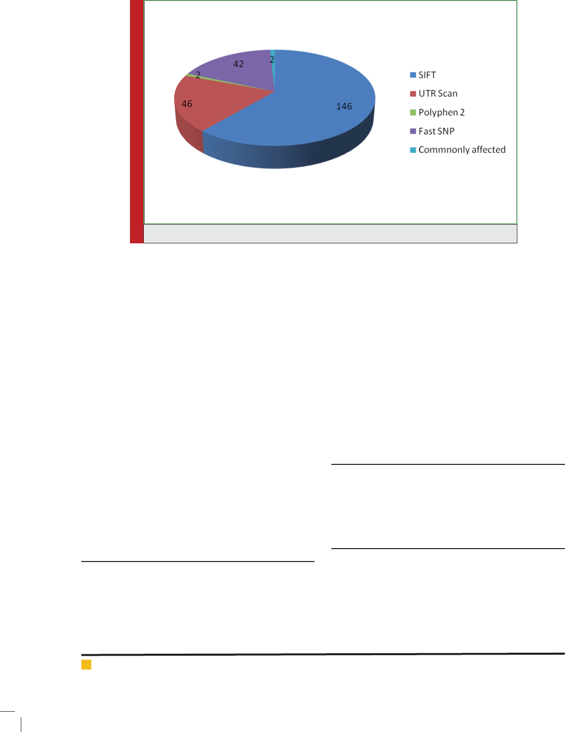
We considered the 64 most potential hindering point
changes for further course of examinations in light of
the fact that they were generally discovered to be less
steady, injurious, and harming by the I-Mutant 3.0, SIFT
and Poly Phen-2 servers individually. The most com-
monly affected among the 6 computational tools has
been taken for further studies i.e. 2 variants as shown
in Figure 3.
CONCLUSION
Hence the combined approach using SIFT, UTRscan and
Polyphen 2 predicts the mutation rs6063 and rs74315205
are most deleterious among the mutations for UCHL1
gene causing Parkinson’s disease characterized. The
recognition of these SNPs as deleterious ones provides
insight into PD biology and presents as anti Parkin-
son’s disease therapeutic targets and diagnostic markers
.Since missense mutations are nucleotide substitutions
that change an amino acid in a protein, the deleterious
effects of these mutations are commonly attributed to
their impact on primary amino acid sequence and pro-
tein structure. Structural analysis results showed that the
amino acid residue substitutions which had the great-
est impact on the stability of the UCHL 1 protein were
mutations in rs6063 and rs74315205 and the variants
like G191R, G199 R, G88R and R231G. Based on our
results, we conclude that these SNPs should be consid-
ered important candidates in UCHL1 related P.D. Based
on our results we conclude that these SNPs should be
considered as important candidates in causing Parkin-
son’s disease.
ACKNOWLEDGEMENT
The authors are highly thankful to the Department of
Biotechnology, Delhi, India for providing support in the
form of Bioinformatics infrastructure facility to carry
out this work.
DISCLOSURE STATEMENT
No competing nancial interests exist.
REFERENCES
Aaron Ciechanover and Yong Tae Kwon (2015). Degradation
of misfolded proteins in neurodegenerative diseases: therapeu-
tic targets and strategies, Experimental & Molecular Medicine.
Volume 47, No.3, pp.1-16.
FIGURE 3. Status of SNPS derived through various algorithms

BIOSCIENCE BIOTECHNOLOGY RESEARCH COMMUNICATIONS IDENTIFICATION OF NOVEL MICRO RNAS AND THEIR TARGETS 511
Sowmya Dhawan and Usha Chouhan
Cargill M D, Altshuler J, Ireland P, Sklar K, Ardlie N,Patil N,
Shaw CR, Lane EP, Lim N, M. Kalyanaraman (1999). Character-
ization of single-nucleotide polymorphisms in coding regions
of human genes Nat. Genet, Volume 23, No.3, pp.373.
Christine Klein and Ana Westenberger A (2012). Genetics of
Parkinson’s disease.Cold Spring Harb Perspective Med,Volume
2, No.1.
Contu VR, Kotake Y, Toyama T, Okuda K, Miyara M, Sakamoto
S, Samizo S, Sanoh S, Kumagai Y, Ohta S (2014). Endogenous
neurotoxic dopamine derivative covalently binds to Parkin-
son’s disease-associated ubiquitin C-terminal hydrolase L1
and alters its structure and function. J Neurochem ,Volume
130,No.6, pp.826-838.
Graziano Pesole and Sabino Liuni ( 1999) .Internet resources
for the functional analysis of 5’ and 3’ untranslated regions
of eukaryotic mRNA,Trends Genet., Volume 15, No.9, pp. 378.
Gómez, J.L Octavio Mercado-Gómez and Rosalinda Guevara-
Guzmán ( 2015) Epigenetic mechanisms in neurological and
neurodegenerative diseases. Frontiers in cellular Neuroscience,
Volume 9, No.58, pp.1-11
Graziano Pesole, Sabino Liuni, G Grillo, F Licciulli, F Mignone,
C Gissi, C Saccone (2002),UTRdb and UTRsite: specialized
databases of sequences and functional elements of 5’ and
3’untranslated regions of eukaryotic mRNAs. Nucleic Acids
Res.,Volume 30, No.1, pp. 335–340.
Hsiang-Yu Y, Jen-Jie C, Wen-Hsien T, Chia-Hung L, Chuan-
Kun L, Yi-Jung L, Hui-Hung W, Adam Y, Yuan-Tsong C, Chun-
Nan H (2006). FASTSNP: an always up-to-date and extendable
service for SNP function analysis and prioritization. Nucleic
Acids Res. Volume .34, W635–W641,pp.635-641.
Hyo Eun Moon and Sun, H P. (2015).Mitochondrial Dysfunc-
tion in Parkinson’s. Experimental neurology. Volume 24, No.2,
pp.103-116.
Kumar P, Henikoff S, Pauline C (2009). Predicting the effects
of coding non-synonymous variants on protein function using
the SIFT algorithm, Nat. Protoc. Volume.4, pp.8-9.
Liu Y, Fallon L, Lashuel HA, Liu Z, Lansbury PT Jr (2002).The
UCH-L1 gene encodes two opposing enzymatic activities that
affect alpha-synuclein degradation and Parkinson’s disease
susceptibility. Cell, Volume 111, No.2, pp.209-218.
Maraganore DM, Lesnick TG, Elbaz A, Chartier-Harlin MC,
Gasser T, Kruger R, Hattori N, Mellick GD, Quattrone A, Satoh
J, Toda T, Wang J, Ioannidis JP, de AM, Rocca WA (2004).
UCHL1 is a Parkinson’s disease susceptibility gene. Ann Neu-
rol.Volume 55, No. 4, pp. 512-521.
Minyue Jia, Boyun Yang, Zhongyi Li, Huiling Shen, Xiaox-
iao Song, Wei Gu (2014) Computational Analysis of Func-
tional Single Nucleotide Polymorphisms Associated with the
CYP11B2 .Plos One, Volume 9, No. 8, pp.1-14.
Paul Bishop, Dan Rocca and Jeremy M. Henley (2016). Ubiq-
uitin C-terminal hydrolase L1 (UCH-L1): structure, distribution
and roles in brain function and dysfunction Biochem. J. Vol-
ume 473, No.16, pp. 2453–2462.
Pauline C Ng, Steven Henikoff (2003), SIFT: predicting amino
acid changes that affect protein function. Nucleic Acids Res,
Volume 31, No.13, pp. 3812–3814.
Pauline C Ng, Steven Henikoff (2001), Predicting deleterious
amino acid substitutions. Genome Res, Volume 11, No. 5, pp.
863–874.
Prasad RKA, Babu SS, Siddaiah N and Rao KS (2016).A Review
on Techniques for Diagnosing and Monitoring Patients with
Parkinson’s disease. Journal of Biosensors & Bioelectronics,
Volume 7. No.2, pp.1-7.
Prokunina L, Alarcn-Riquelme ME (2004). Regulatory SNPs in
complex diseases: their identi cation and functional valida-
tion. Expert Rev. Mol. Med.Volume 6, No. 10, pp. 1–15.
Prokunina L, Castillejo-Lopez C,Oberg F, Gunnarsson I, Berg L,
Magnusson V, Brookes AJ, Tentler AJ, Kristjansdottir H, Gron-
dal G, et al.(2002). A regulatory polymorphism inPDCD1 is
associated with susceptibility to systemic lupus erythematosus
in humans. Nat. Genet., Volume 32, No. 4, pp. 666–669.
Ramensky V, Bork, P, Sunyaev S,(2002). Human non-synony-
mous SNPs: server and survey. Nucleic Acids Res. Volume .30,
No.17, pp. 3894–3900.
Smith K (2002). Genetic Polymorphism and SNPs Genotyp-
ing, Haplotype Assembly Problem Haplotype Map.Functional
Genomics and Proteomics.
Sunyaev S, Ramensky V, Bork P (2000), Towards a struc-
tural basis of human non synonymous single nucleotide
polymorphisms,Trends Genet, Volume 16, No. 5, pp. 198–200.